Anatomical Drawing Of The Brain
Anatomical Drawing Of The Brain - Figures of the brain appear to be done after external fixation in the work of willis. Web to draw an anatomically accurate brain, draw a curve in the shape of the lengthwise half of a large egg, making the right side more curved. Illustrations and diagrams of the brain These works reflected his efforts to understand medieval psychology, including the localisation of sensory and motor functions to the brain. It sits mainly in the anterior and middle cranial fossae of the skull. Drawn mainly from the collections of the nlm, dream anatomy shows off the anatomical imagination in some of its most astonishing incarnations, from 1500 to the present. The brain illustrations of vesalius and willis were the first in anatomic history with pictorial accuracy. Web the brain illustrations of vesalius and willis were the first in anatomic history with pictorial accuracy. Web the lateral view of the brain shows the three major parts of the brain: Together, the brain and spinal cord that extends from it make up the central nervous system, or cns. Together, the brain and spinal cord that extends from it make up the central nervous system, or cns. Their illustrations, illustrators, and methods are discussed. This will help with your exams and can score higher marks. See brain anatomical drawing stock video clips. The text accompanying the drawing also attributes the brain with imaginativa, logistica and memoria, thereby showing knowledge. The cerebrum is the largest and most recognizable part of the brain. Web “cells of the brain” presents some of the basics, beginning with pyramidal neurons, and including the pericellular nests that surround them like pointy hats, or eva hesse sculpture, and. Their illustrations, illustrators, and methods are discussed. Utilize anatomy books, online resources, or 3d models to study the. Their illustrations, illustrators, and methods are discussed. Drawn mainly from the collections of the nlm, dream anatomy shows off the anatomical imagination in some of its most astonishing incarnations, from 1500 to the present. Web the brain is a complex organ that controls thought, memory, emotion, touch, motor skills, vision, breathing, temperature, hunger and every process that regulates our body.. Woodcut blocks were used for the prints of figures in the vesalian anatomy. Web the brain illustrations of vesalius and willis were the first in anatomic history with pictorial accuracy. Web the brain is composed of the cerebrum, cerebellum, and brainstem (fig. The brain illustrations of vesalius and willis were the first in anatomic history with pictorial accuracy. Axial mri. It is composed of 64 drawings, illustrations and anatomical charts, all in vector format. Their illustrations, illustrators, and methods are discussed. A lateral view of the cerebrum is the best perspective to appreciate the lobes of the hemispheres. Anatomical structure of the head and neck. All images photos vectors illustrations 3d objects. Their illustrations, illustrators, and methods are discussed. Web at present, it is known that the brain has an anatomical and functional distribution due to the complexity of the organization of the cells. Web “cells of the brain” presents some of the basics, beginning with pyramidal neurons, and including the pericellular nests that surround them like pointy hats, or eva hesse. While drawing from imagination is a valuable skill, having reference images of the brain's anatomy can provide valuable guidance and ensure anatomical accuracy in your illustration. Web basic anatomy and function of the brain. Web brain anatomy drawing stock photos are available in a variety of sizes and formats to fit your needs. Web the lateral view of the brain. Human brain sagittal view medical sketchy illustration. Reviewed by john morrison, patrick hof, and edward lein. While drawing from imagination is a valuable skill, having reference images of the brain's anatomy can provide valuable guidance and ensure anatomical accuracy in your illustration. A lateral view of the cerebrum is the best perspective to appreciate the lobes of the hemispheres. Web. This division due to the cortex organization of highly compacted neurons. Click on the bodymap above to interact with a 3d model of the brain. It consists of grey matter (the cerebral cortex) and white matter at the center. Figures of the brain appear to be done after external fixation in the work of willis. See brain anatomical drawing stock. Web basic anatomy and function of the brain. The brain illustrations of vesalius and willis were the first in anatomic history with pictorial accuracy. This part of the brain is. Click on the bodymap above to interact with a 3d model of the brain. Reviewed by john morrison, patrick hof, and edward lein. It consists of grey matter (the cerebral cortex) and white matter at the center. Human brain sagittal view medical sketchy illustration. See brain anatomical drawing stock video clips. Woodcut blocks were used for the prints of figures in the vesalian anatomy. The cerebrum is the largest and most recognizable part of the brain. The human brain consists of several parts which are clearly labeled in the video. Utilize anatomy books, online resources, or 3d models to study the structure of the brain. Woodcut blocks were used for the prints of figures in the vesalian anatomy. Web anatomy of the brain: Web “cells of the brain” presents some of the basics, beginning with pyramidal neurons, and including the pericellular nests that surround them like pointy hats, or eva hesse sculpture, and. Each hemisphere is conventionally divided into six lobes, but only four of them are visible from this lateral perspective. Hand drawn line art anatomically correct human brain. It is composed of 64 drawings, illustrations and anatomical charts, all in vector format. Click on the bodymap above to interact with a 3d model of the brain. The brain has three main parts: The cerebrum, also called the telencephalon, refers to the two cerebral hemispheres (right and left) which form the largest part of the brain.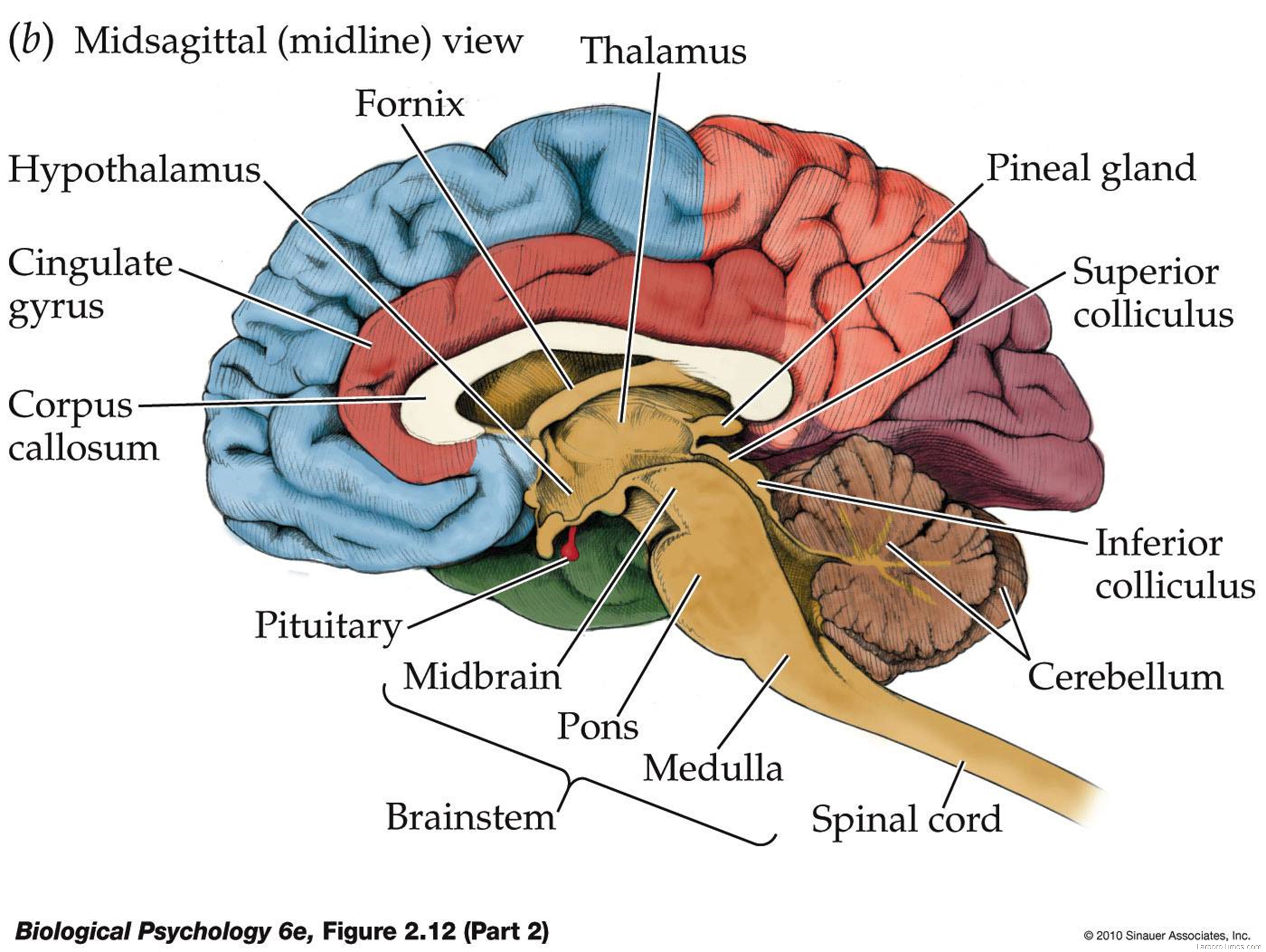
brain diagram Anatomy System Human Body Anatomy diagram and chart
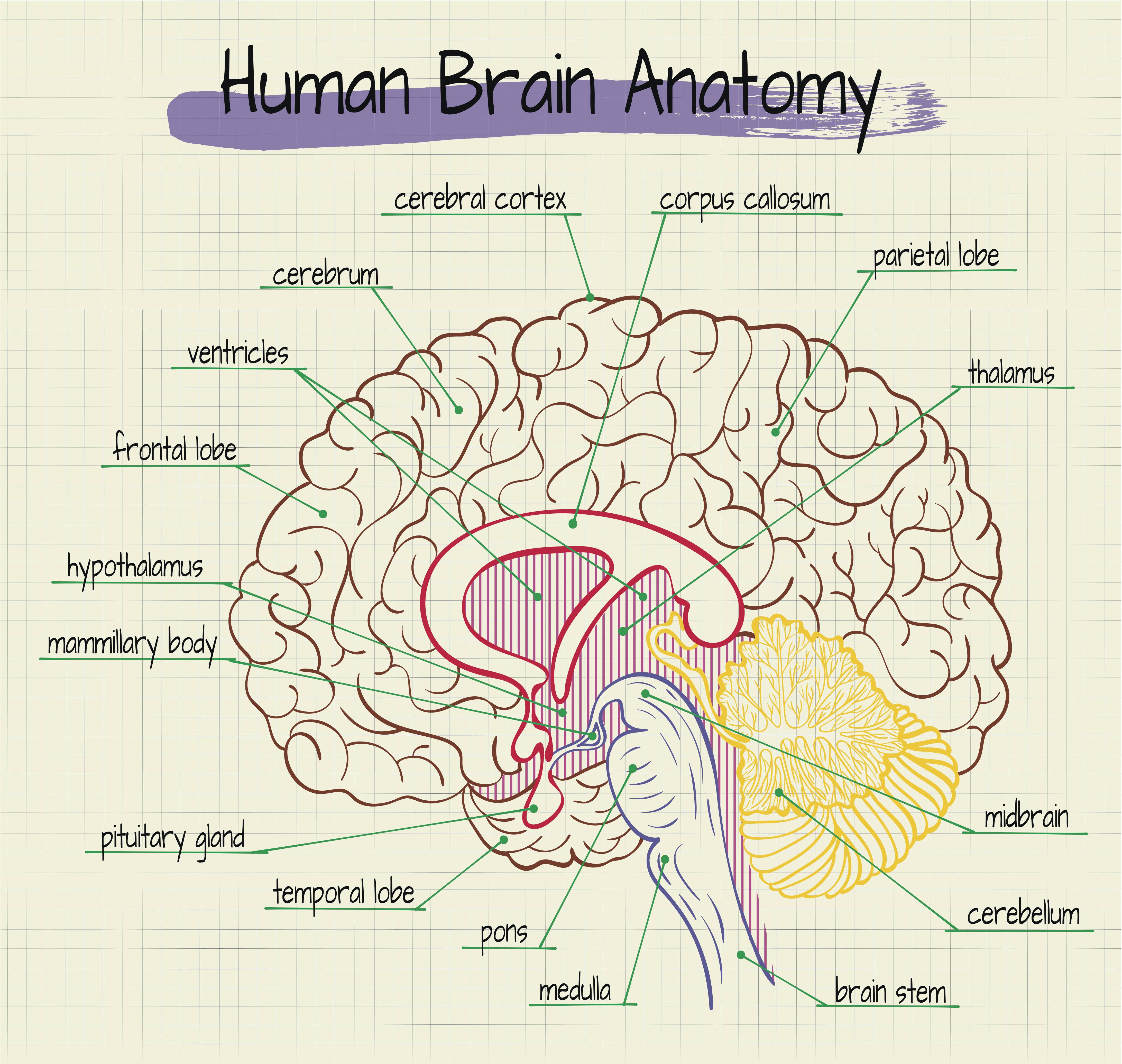
brainanatomy Rachel Gold
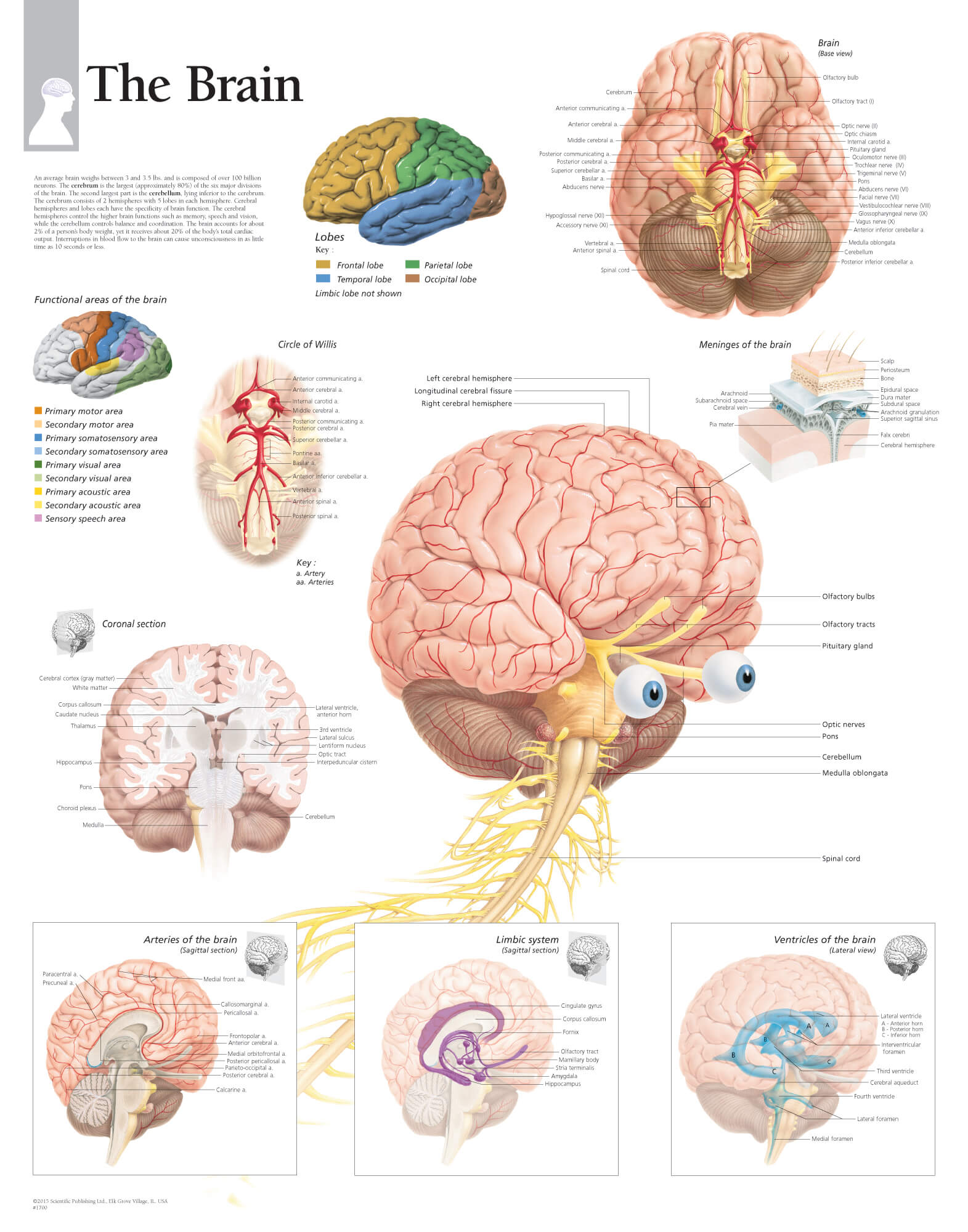
The Brain Scientific Publishing
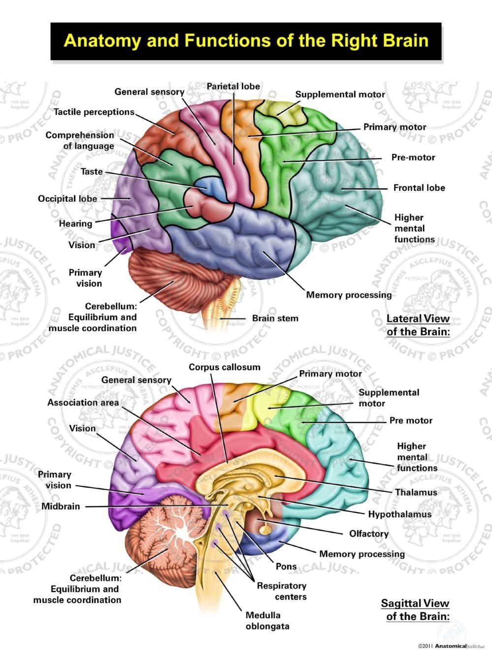
Anatomy of the Human Brain Essentiality of More Indepth Studies The
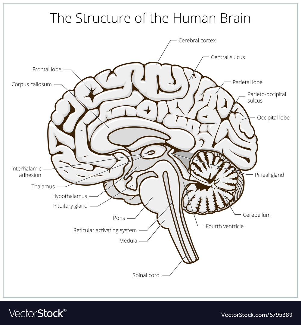
Structure of human brain section schematic Vector Image
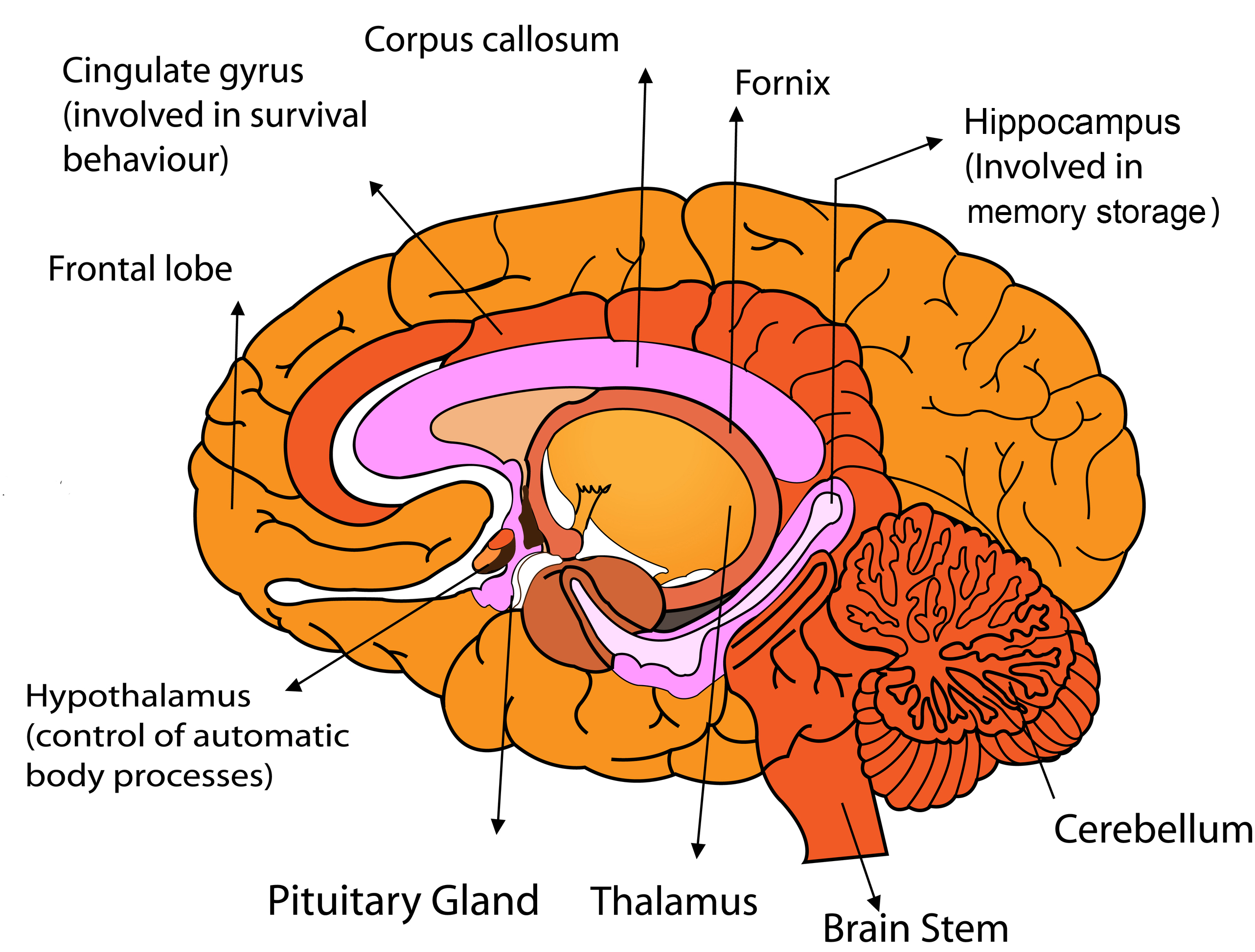
Parts of the Human Brain
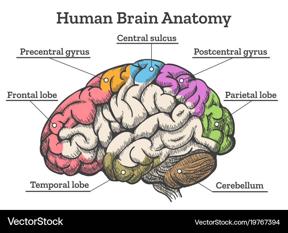
Human brain anatomy diagram Royalty Free Vector Image

How to Draw a Brain 14 Steps wikiHow
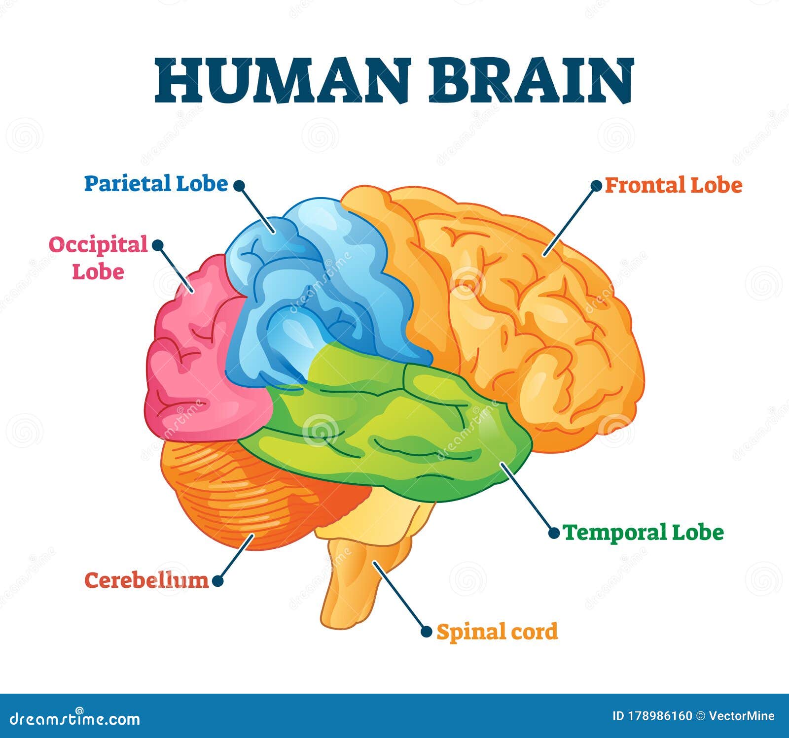
Human Brain Vector Illustration. Labeled Anatomical Educational Parts

Diagram of Human Brain System Health Images Reference
Print Or Tattoo Design Isolated On White Background Vector Illustration.
Drawn Mainly From The Collections Of The Nlm, Dream Anatomy Shows Off The Anatomical Imagination In Some Of Its Most Astonishing Incarnations, From 1500 To The Present.
Anatomical Structure Of The Head And Neck.
Web At Present, It Is Known That The Brain Has An Anatomical And Functional Distribution Due To The Complexity Of The Organization Of The Cells.
Related Post: