Bronchioles Drawing
Bronchioles Drawing - The trachea branches into the. The bronchioles typically begin beyond the tertiary segmental bronchi and are described as conducting bronchioles. Each alveolar duct has 5 or 6 associated alveolar sacs. Web how to draw the human respiratory system. Conduct air but they lack glands or alveoli; Bronchioles and alveoli histology videos, flashcards, high yield notes, & practice questions. 2 adding accessory structures and details. Bronchioles are small tubes that branch off of bronchi and connect to the air sacs (alveoli) in the lungs. Web perhaps start with the left lung, which would be on the right side, first drawing the bronchioles. Web human anatomy laboratory manual 2021. Web how to draw the human respiratory system. The primary bronchi, in each lung, which are the left and right bronchus, give rise to secondary bronchi. Web the bronchioles are air passages in the lungs that deliver air to the alveoli. These bronchioles give rise to alveolar ducts that then give rise to alveolar sacs. Watch the video tutorial now. Web a bronchus (plural bronchi, adjective bronchial) is a passage of airway in the respiratory tract that conducts air into the lungs. Web what are bronchioles definition, where are they located, description, anatomy (terminal, respiratory bronchioles), what do bronchioles do in respiratory system. The word is derived from the greek word bronchos, which refers to. Learn and reinforce your understanding. These bronchioles give rise to alveolar ducts that then give rise to alveolar sacs. These in turn give rise to tertiary bronchi (tertiary meaning third). The bronchus branches into smaller tubes called bronchioles. Web respiratory bronchioles are lined with simple cuboidal epithelium. The alveolus is the basic anatomic unit of gas exchange. Web the segmental bronchi divide into many smaller bronchioles that divide into terminal bronchioles, and then into respiratory bronchioles, which divide into 2 to 11 alveolar ducts. Bronchioles are small tubes that branch off of bronchi and connect to the air sacs (alveoli) in the lungs. Each alveolar duct has 5 or 6 associated alveolar sacs. Web the bronchioles are. Each respiratory bronchiole branches to a few alveolar ducts that connect to alveolar sacs in the respiratory zone. Staying with the left lung, once you have drawn the various bronchial branches, proceed to draw the various lines. Each terminal bronchiole branches to several respiratory bronchioles. Watch the video tutorial now. Web this is our sneak peek at the full tutorial. Each respiratory bronchiole branches to a few alveolar ducts that connect to alveolar sacs in the respiratory zone. The trachea branches into the. Web respiratory bronchioles are lined with simple cuboidal epithelium. Drawing shows the right lung with the upper, middle, and lower lobes, the left lung with the upper and lower lobes, and the trachea, bronchi, lymph nodes, and. Web the bronchioles are air passages in the lungs that deliver air to the alveoli. Bronchioles and alveoli histology videos, flashcards, high yield notes, & practice questions. These bronchioles give rise to alveolar ducts that then give rise to alveolar sacs. The bronchioles are tubes in the lungs which branch off from the larger bronchi that enter each lung, from. Conduct air and also contain alevoli that extend from their lumens. The word is derived from the greek word bronchos, which refers to. The bronchioles typically begin beyond the tertiary segmental bronchi and are described as conducting bronchioles. Web perhaps start with the left lung, which would be on the right side, first drawing the bronchioles. Web how to draw. Web this is our sneak peek at the full tutorial about the bronchioles and alveoli. The primary bronchi, in each lung, which are the left and right bronchus, give rise to secondary bronchi. Web there are two types of bronchioles: These in turn give rise to tertiary bronchi (tertiary meaning third). Bronchioles are small tubes that branch off of bronchi. By following the simple steps, you too can easily draw a perfect lungs. These bronchioles give rise to alveolar ducts that then give rise to alveolar sacs. The trachea branches into the. The alveolus is the basic anatomic unit of gas exchange. Web bronchioles are the branches of the tracheobronchial tree that by definition, are lacking in submucosal hyaline cartilage. Infection with bacteria or viruses can also lead to conditions like bronchopneumonia. Learn the fine structure of the lungs. Web there are two types of bronchioles: Learn and reinforce your understanding of bronchioles and alveoli histology. Web bronchioles are the branches of the tracheobronchial tree that by definition, are lacking in submucosal hyaline cartilage. Conduct air and also contain alevoli that extend from their lumens. They can be affected by a number of different conditions, from asthma to cystic fibrosis. These bronchioles give rise to alveolar ducts that then give rise to alveolar sacs. Web perhaps start with the left lung, which would be on the right side, first drawing the bronchioles. The tertiary bronchi subdivide into the bronchioles (respiratory bronchioles). Conduct air but they lack glands or alveoli; Web what are bronchioles definition, where are they located, description, anatomy (terminal, respiratory bronchioles), what do bronchioles do in respiratory system. It differs from the other bronchioles in that its walls contain alveoli, in which gas exchange occurs. Web so here's a little person, and i've drawn their face, and you can see in blue at the bottom i've drawn their voice box, and i'm going to show you exactly what happens when you draw a little molecule like this of oxygen and to follow it along it's journey. Web this is our sneak peek at the full tutorial about the bronchioles and alveoli. 1 drawing basic respiratory structures.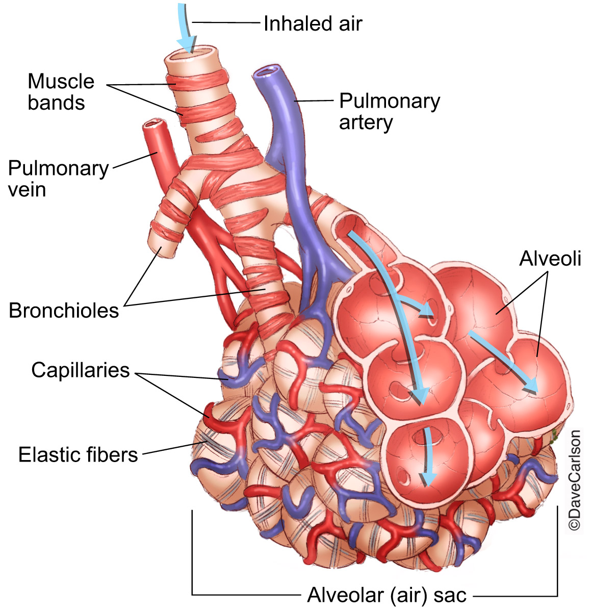
Lung Bronchioles & Alveoli Carlson Stock Art
![]()
Human lungs with bronchi and bronchioles linear icon. Thin line
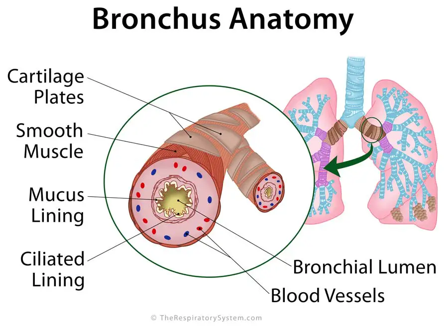
Bronchi Definition, Location, Anatomy, Functions, Pictures
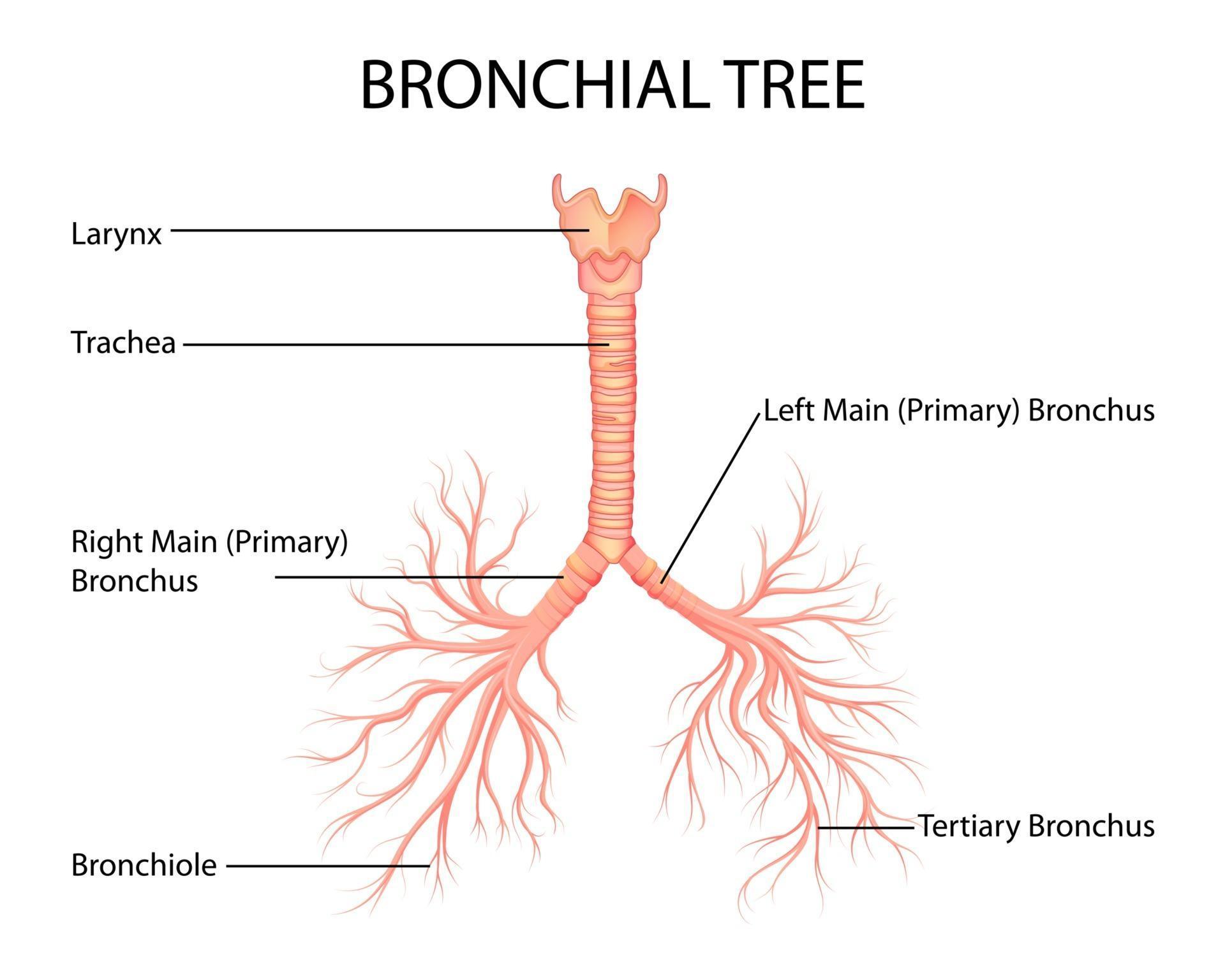
illustration of Healthcare and Medical education drawing chart of Human
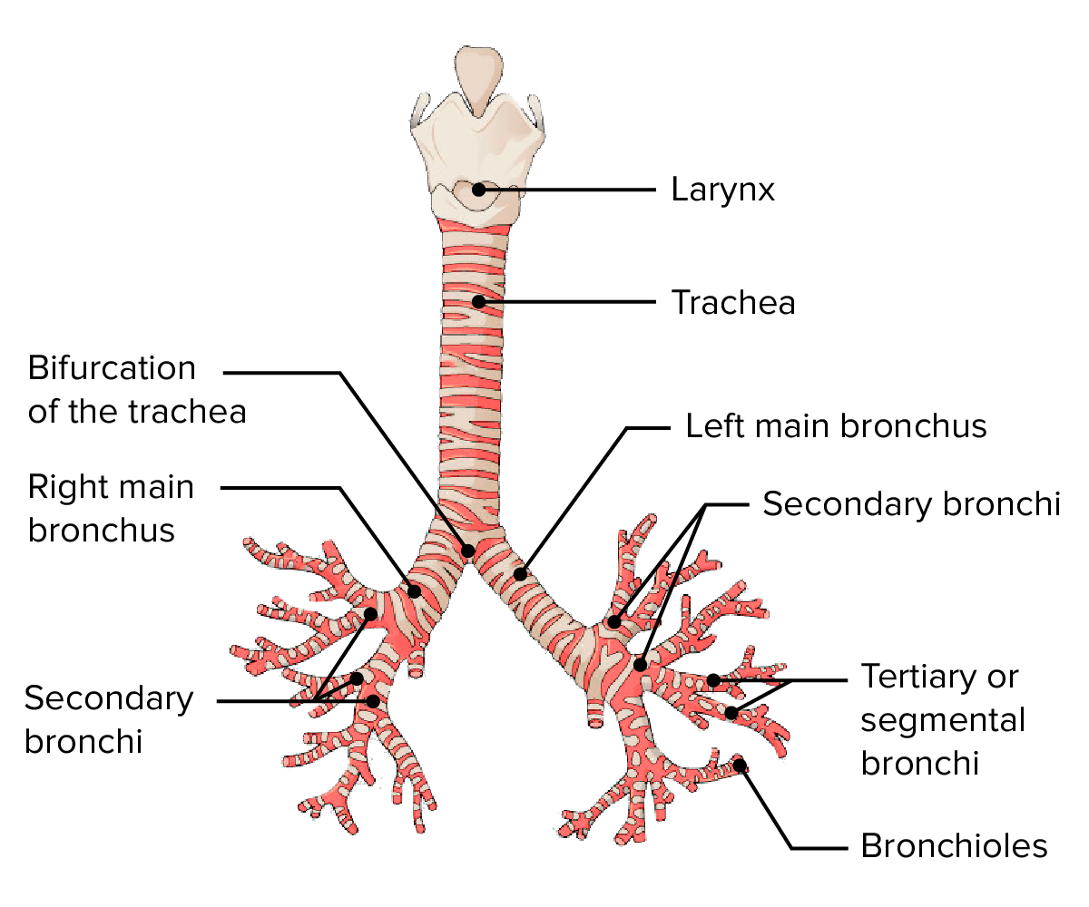
Human Respiratory System Diagram Class 10 CBSE Class Notes Online

NROER File, Image Structure of Affected Bronchioles
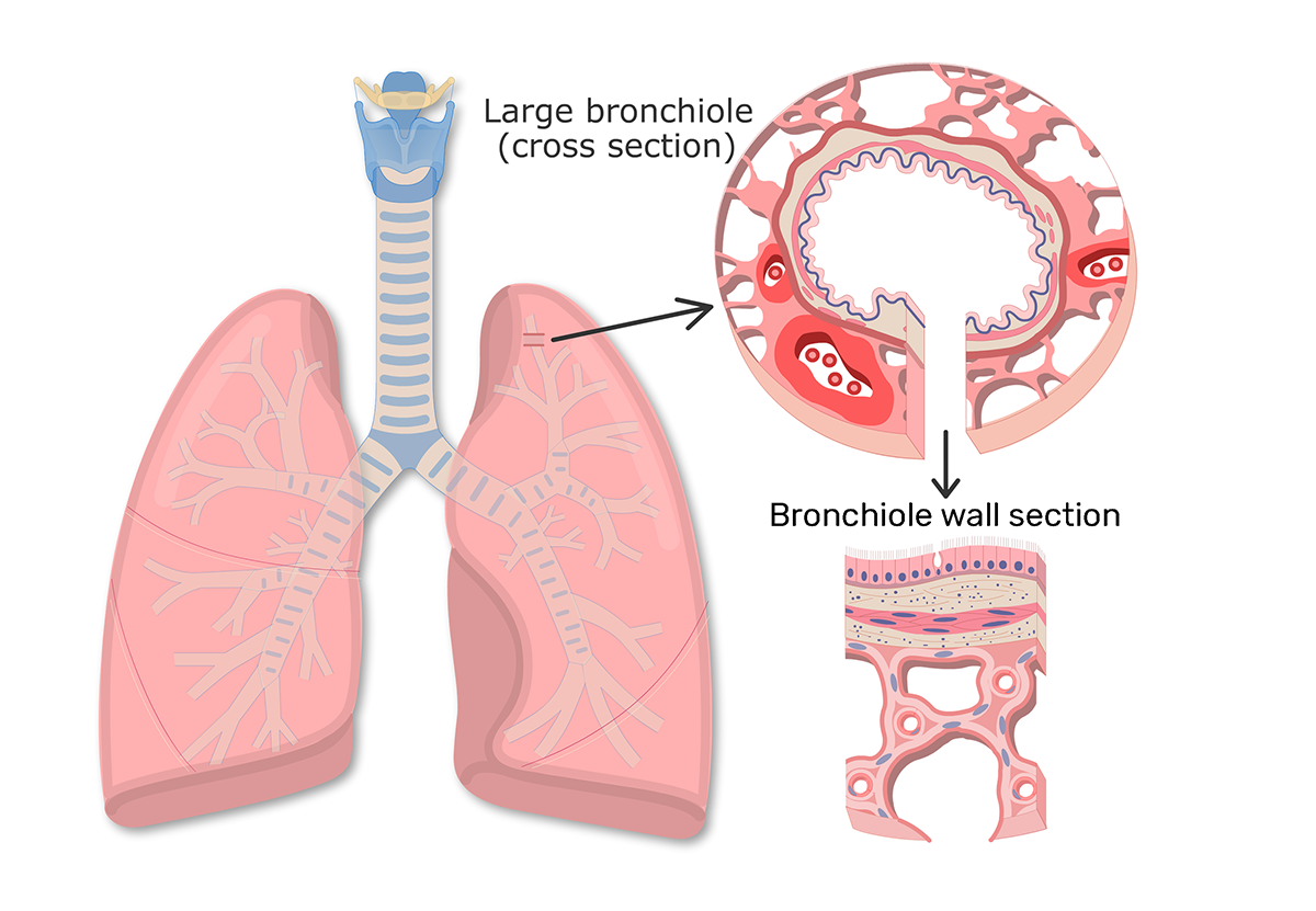
Bronchioles function and diagram GetBodySmart
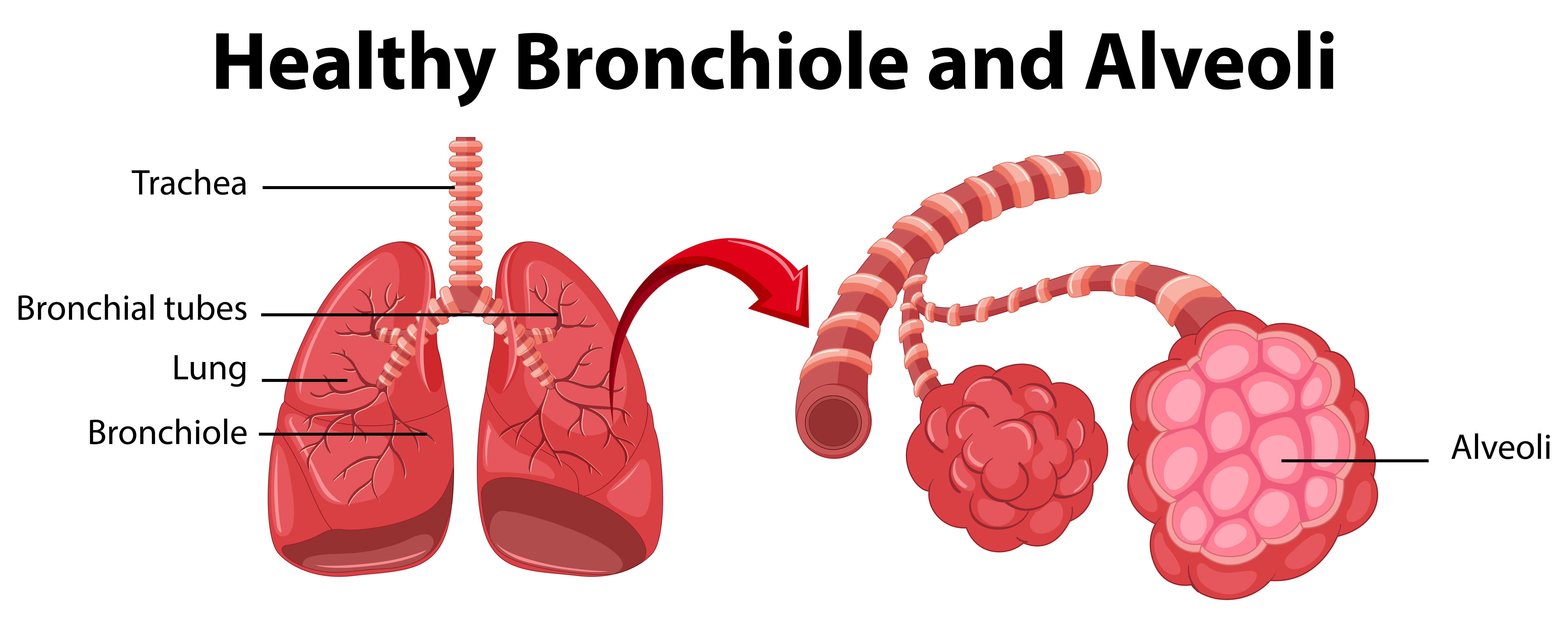
Diagram showing healthy bronchiole and alveoli 434375 Vector Art at

Anatomy of the bronchus and bronchial tubes Stock Photo Alamy
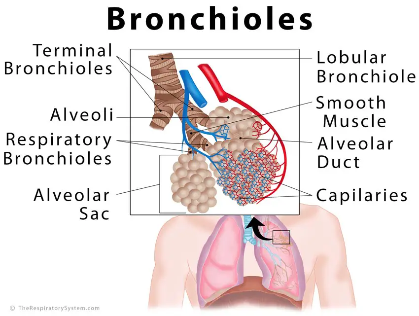
Bronchioles Diagram
Begin At The Bifurcation Of The Trachea At The Sternal Angle (T4/T5) And Enters The Hilum Of Each Lung.
Each Alveolar Duct Has 5 Or 6 Associated Alveolar Sacs.
Web Image Collection Gallery List.
Watch The Video Tutorial Now.
Related Post: