Bronchus Drawing
Bronchus Drawing - There are several medical illustration standards to keep in mind, such as using blue for oxygenated blood flow and red for deoxygenated blood flow. A crucial part of the respiratory system, the bronchi function primarily as passageways for air, bringing oxygen. Web this is our sneak peek at the full tutorial about the bronchioles and alveoli. You can see that the major bronchi are visible if you look carefully. All images photos vectors illustrations 3d objects. Bronchi will branch into smaller tubes that become bronchioles. Web the lungs are most often considered as part of the lower respiratory tract, but are sometimes described as a separate entity. At the end of the bronchioles are air sacs called alveoli , and this is where gas exchange occurs. Diagram labeling the major structures of the respiratory system Web human lungs drawing is a fun exercise due to the strange visual nature of the lungs. Web drawing basic respiratory structures. Web this is our sneak peek at the full tutorial about the bronchioles and alveoli. Illustration of a man's bronchi. This article will discuss the anatomy and function of the respiratory system. The trachea (windpipe) is found inferior to the thyroid cartilage and superior to division into the left and right main bronchus. Human trachea and bronchi, larynx. Web the bronchi are the two large tubes that carry air from the windpipe into the lungs and back out again. Illustration of a man's bronchi. It may be beneficial to practice drawing the bronchi and labeling them until you are entirely familiar with their names and. At the end of the bronchioles are air. Asthma disease vector icon in flat style. 22.1 x 40.1 cm ⏐ 8.7 x 15.8 in (300dpi) this image is not available for purchase in your country. After the bronchi, remember that the left pulmonary artery arches over the left upper lobe bronchus and the right pulmonary artery passes posterior to the ascending aorta to divide into the truncus anterior. Web each bronchus enters the root of the lung, passing through the hilum. Human respiratory system anatomical poster. Each lobar bronchus then further divides into several tertiary segmental bronchi. Web so here's a little person, and i've drawn their face, and you can see in blue at the bottom i've drawn their voice box, and i'm going to show you. Bronchi will branch into smaller tubes that become bronchioles. The direction of the bronchi advancing the periphery is close to orthogonal to the ct section. They contain the respiratory bronchioles, alveolar ducts, alveolar sacs and alveoli. Request price add to basket remove add to board. Diagram labeling the major structures of the respiratory system Web so here's a little person, and i've drawn their face, and you can see in blue at the bottom i've drawn their voice box, and i'm going to show you exactly what happens when you draw a little molecule like this of oxygen and to follow it along it's journey. Each lobar bronchus then further divides into several tertiary. Please contact your account manager if you have any query. Asthma disease vector icon in flat style. Web each bronchus divides into smaller bronchi, and again into even smaller tubes called bronchioles. The following schematic drawing should help you sort out these structures. It may be beneficial to practice drawing the bronchi and labeling them until you are entirely familiar. Web so here's a little person, and i've drawn their face, and you can see in blue at the bottom i've drawn their voice box, and i'm going to show you exactly what happens when you draw a little molecule like this of oxygen and to follow it along it's journey. Web your sketch should include the nasal cavity, the. Rings and left main bronchi. Bronchi will branch into smaller tubes that become bronchioles. Illustration of a man's bronchi. Web each bronchus divides into smaller bronchi, and again into even smaller tubes called bronchioles. Web this is our sneak peek at the full tutorial about the bronchioles and alveoli. Web each bronchus enters the root of the lung, passing through the hilum. The lung’s strange composition is a fun and interesting subject to draw as the lungs are seemingly alien and abstract. This article will discuss the anatomy and function of the respiratory system. Web four patterns of tracing the bronchus, based on the direction of the bronchial branch. 35.2 mb (507.6 kb compressed) 2601 x 4733 pixels. All images photos vectors illustrations 3d objects. Web in this first step of our guide on how to draw lungs, we will be drawing the central structure of the lungs, called the trachea. Each lobar bronchus then further divides into several tertiary segmental bronchi. Web choose from 354 drawing of bronchial tubes stock illustrations from istock. Begin at the bifurcation of the trachea at the sternal angle (t4/t5) and enters the hilum of each lung. Pulmonary alveoli at the end of the bronchioles. The lung’s strange composition is a fun and interesting subject to draw as the lungs are seemingly alien and abstract. Web the lungs are most often considered as part of the lower respiratory tract, but are sometimes described as a separate entity. Human respiratory system anatomical poster. Rings and left main bronchi. At the end of the bronchioles are air sacs called alveoli , and this is where gas exchange occurs. In this posterior view, the left lobar and segmental bronchi are depicted. The trachea (windpipe) is found inferior to the thyroid cartilage and superior to division into the left and right main bronchus. Bronchi will branch into smaller tubes that become bronchioles. Web drawing basic respiratory structures.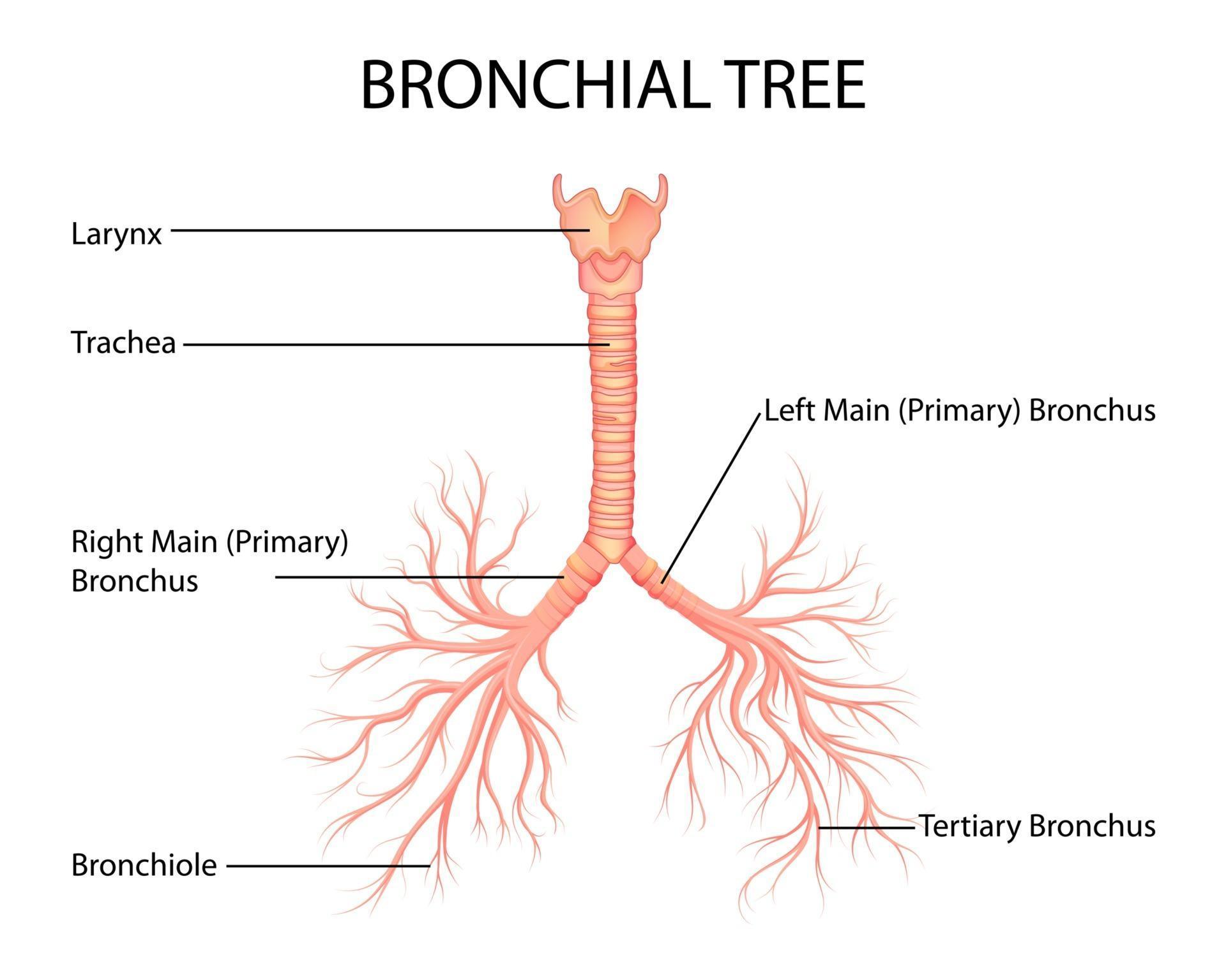
illustration of Healthcare and Medical education drawing chart of Human
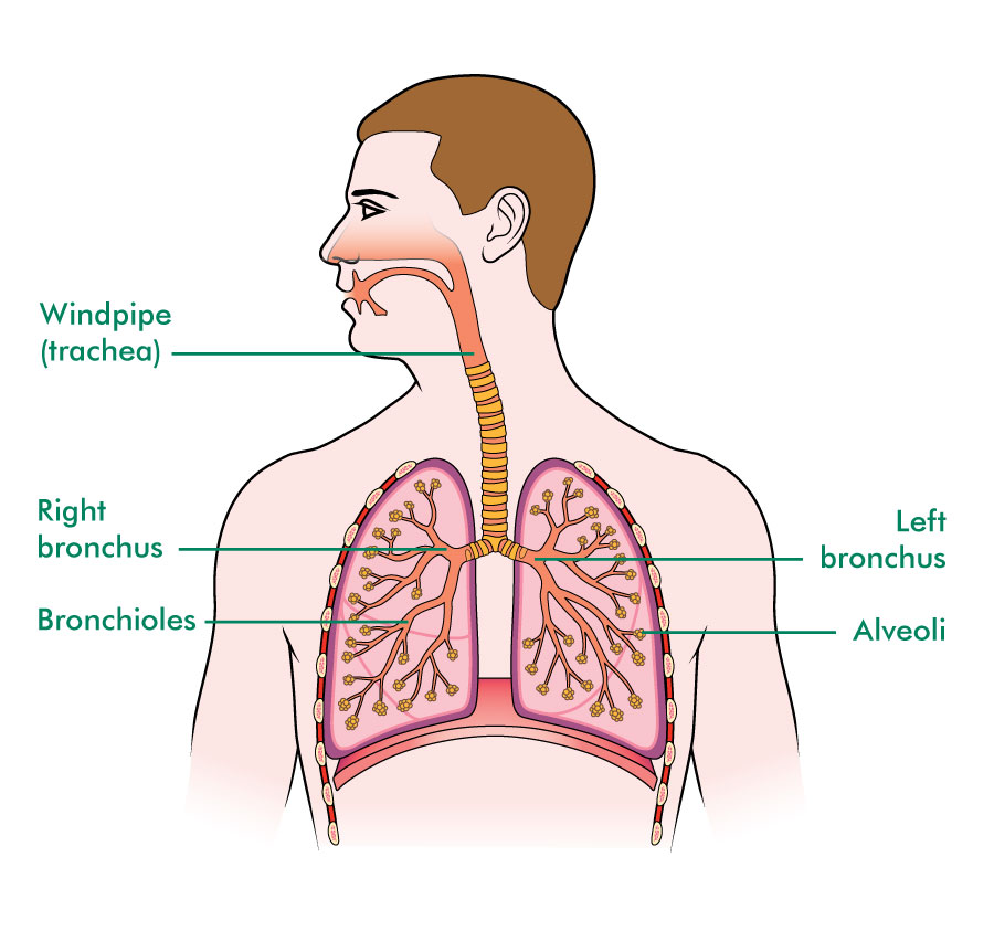
Anatomy Of Bronchioles Anatomical Charts & Posters

BRONCHUS, DRAWING Stock Photo Alamy
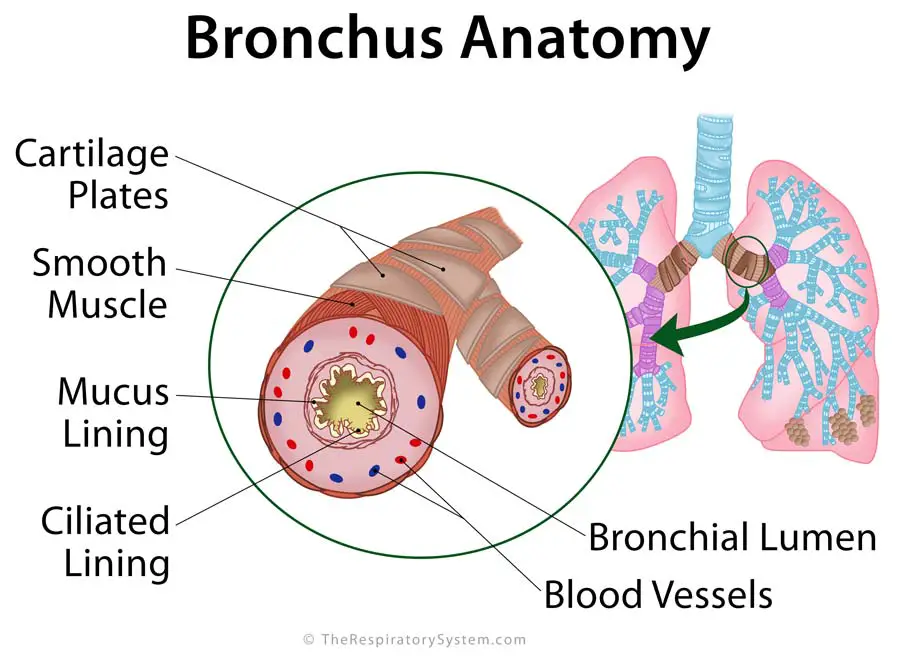
Bronchi Definition, Location, Anatomy, Functions, Pictures
![]()
Human lungs with bronchi and bronchioles linear icon. Thin line
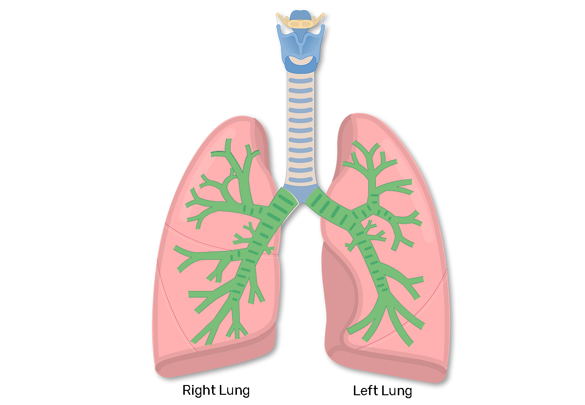
Bronchial Tubes Structure, Functions, & Location Bronchus Anatomy

How to Draw Lungs Really Easy Drawing Tutorial
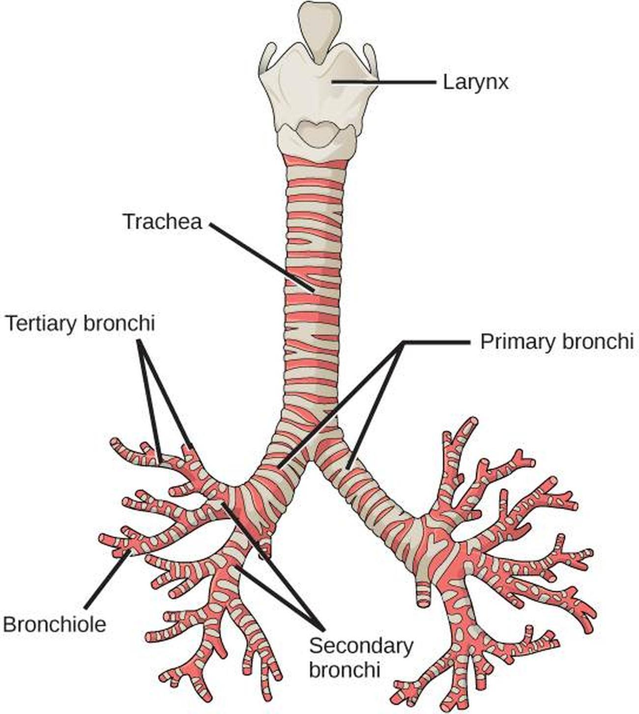
Pictures Of Bronchi

Anatomy of the bronchus and bronchial tubes Stock Photo Alamy
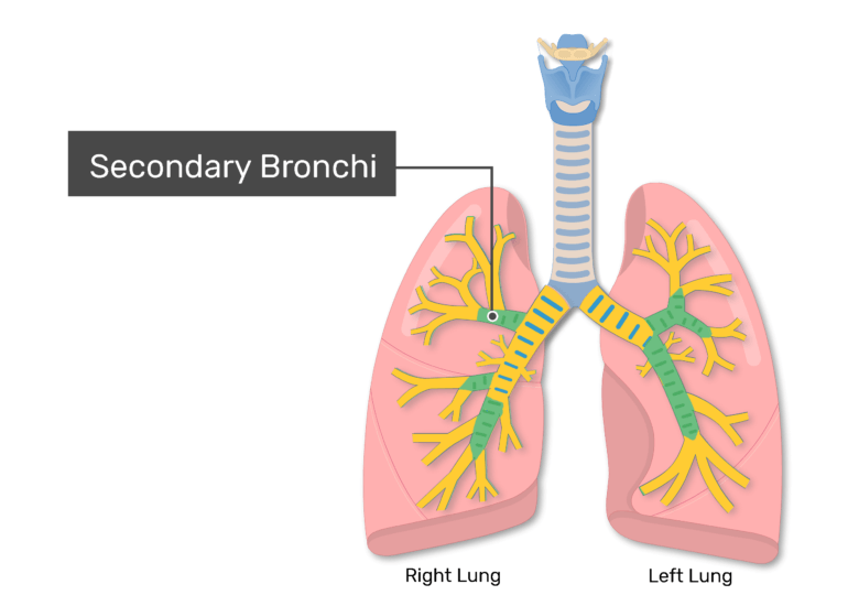
Bronchial Tubes Structure, Functions, & Location Bronchus Anatomy
22.1 X 40.1 Cm ⏐ 8.7 X 15.8 In (300Dpi) This Image Is Not Available For Purchase In Your Country.
Left Bronchial Tree From Posterior.
Case Courtesy Of Dr Matt Skalski, Radiopaedia.org.
There Are Several Medical Illustration Standards To Keep In Mind, Such As Using Blue For Oxygenated Blood Flow And Red For Deoxygenated Blood Flow.
Related Post: