Draw The Stages Of Meiosis
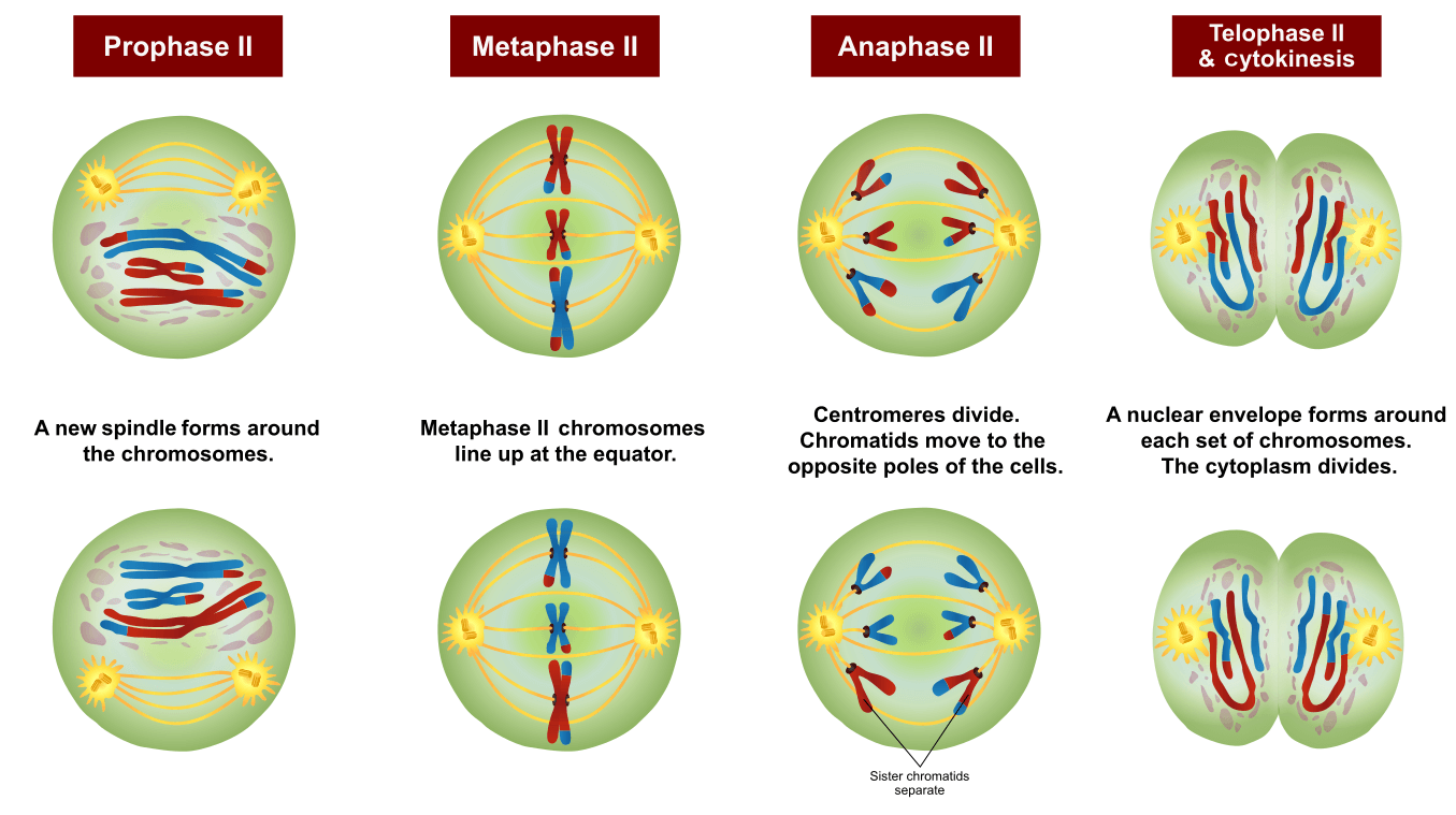
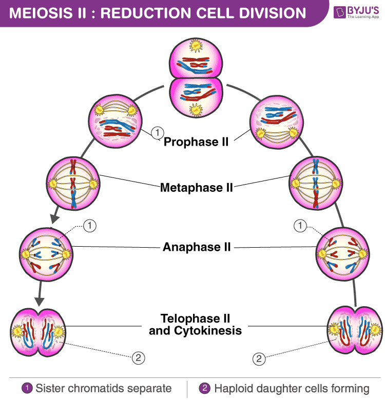
Draw The Stages Of Meiosis - Describe cellular events during meiosis. Web understanding the steps of meiosis is essential to learning how errors occur. Prophase, metaphase, anaphase, and telophase. Web there are six stages within each of the divisions, namely prophase, prometaphase, metaphase, anaphase, telophase and cytokinesis. During prophase i, chromosomes pair up and exchange genetic material, creating more variation. Web describe and draw the key events and stages of meiosis that lead to haploid gametes. Although not a part of meiosis, the cells before entering meiosis i undergo a compulsory growth period called interphase. Meiosis i and meiosis ii. Web here’s a breakdown of the stages of meiosis and a look at what happens: Prophase i is longer than the mitotic prophase and is further subdivided into 5 substages, leptotene. Web there are two stages or phases of meiosis: Web describe and draw the key events and stages of meiosis that lead to haploid gametes. Prophase i, metaphase i, anaphase i, and telophase i. Web prophase i is divided into five different stages: I am demonstrating the colorful diagram of meiosis / phases of meiosis (cell. Before entering meiosis i, a cell must first go through interphase. Web prophase i is divided into five different stages: Before a dividing cell enters meiosis, it undergoes a period of growth called interphase. The sister chromatids remain attached to each other. Each stage includes a period of nuclear division or karyokinesis and a cytoplasmic division or cytokinesis. Cells enter meiosis from interphase, which is much like interphase in mitosis (the cell cycle ). Describe the behavior of chromosomes during meiosis. Explain the mechanisms within meiosis that generate genetic variation among the products of meiosis. In each round of division, cells go through four stages: How to draw the stages of meiosis in exam is the topic. Before entering meiosis i, a cell must first go through interphase. Web meiosis involves two successive stages or phases of cell division, meiosis i and meiosis ii. Web in meiosis i, cells go through four phases: 15k views 4 years ago science diagrams | explained and labelled science diagrams. Although not a part of meiosis, the cells before entering meiosis. The g 1 phase (the “first gap phase”) is focused on cell growth. Each pair of chromosomes—called a tetrad, or a bivalent—consists of four chromatids. Before entering meiosis i, a cell must first go through interphase. The ability to reproduce in kind is a basic characteristic of all living things. The sister chromatids remain attached to each other. In this article, we will look at the stages of meiosis and consider its significance in disease. Web meiosis cell division takes place in the following stages: Remember, before meiosis starts the normally diploid dna has been duplicated. Meiosis is a process where germ cells divide to produce gametes, such as sperm and egg cells. The sister chromatids remain attached. Web describe and draw the key events and stages of meiosis that lead to haploid gametes. Web in each round of division, cells go through four stages: 15k views 4 years ago science diagrams | explained and labelled science diagrams. Meiosis i and meiosis ii. Web meiosis cell division takes place in the following stages: Meiosis i and meiosis ii. Before entering meiosis i, a cell must first go through interphase. Before a dividing cell enters meiosis, it undergoes a period of growth called interphase. Web stages/phases of meiosis. The period prior to the synthesis of dna. Web meiosis involves only one round of dna replication where each chromosome replicates to form sister chromatids. I am demonstrating the colorful diagram of meiosis / phases of meiosis (cell. In this article, we will look at the stages of meiosis and consider its significance in disease. Web there are two stages or phases of meiosis: Web in meiosis i,. When cells commit to meiosis, dna replicates. In kind means that the offspring of any organism closely resemble their parent or parents. The homologous chromosomes are pulled on the opposite poles. Web meiosis begins with prophase i and the contraction of the chromosomes in the nucleus of the diploid cell. Web meiosis involves two successive stages or phases of cell. Prophase i, metaphase i, anaphase i, and telophase i. Prophase i is longer than the mitotic prophase and is further subdivided into 5 substages, leptotene. As in mitosis, the cell grows during g 1 phase, copies all of its chromosomes during s phase, and prepares for division during g 2 phase. Describe cellular events during meiosis. In humans, there are 46 chromosomes or 46 pairs of chromatids. Web prophase i is divided into five different stages: Meiosis consists of two cell divisions namely meiosis 1 and meiosis 2. Each stage includes a period of nuclear division or karyokinesis and a cytoplasmic division or cytokinesis. Organisms that reproduce sexually are thought to have an advantage over. Web there are two stages or phases of meiosis: Prophase, metaphase, anaphase, and telophase. Before entering meiosis i, a cell must first go through interphase. How to draw the stages of meiosis in exam is the topic. Homologous paternal and maternal chromosomes pair up along the midline of the cell. Therefore, when meiosis is completed, each daughter cell contains only half the number (n) of chromosomes as the original cell. Remember, before meiosis starts the normally diploid dna has been duplicated.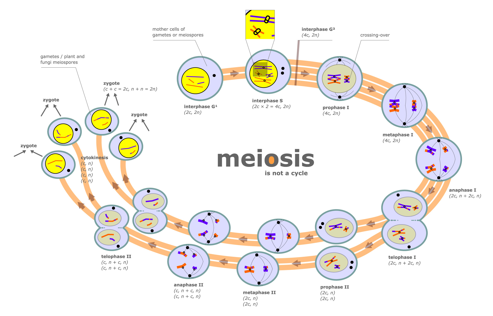
FileMeiosis diagram.jpg Wikipedia
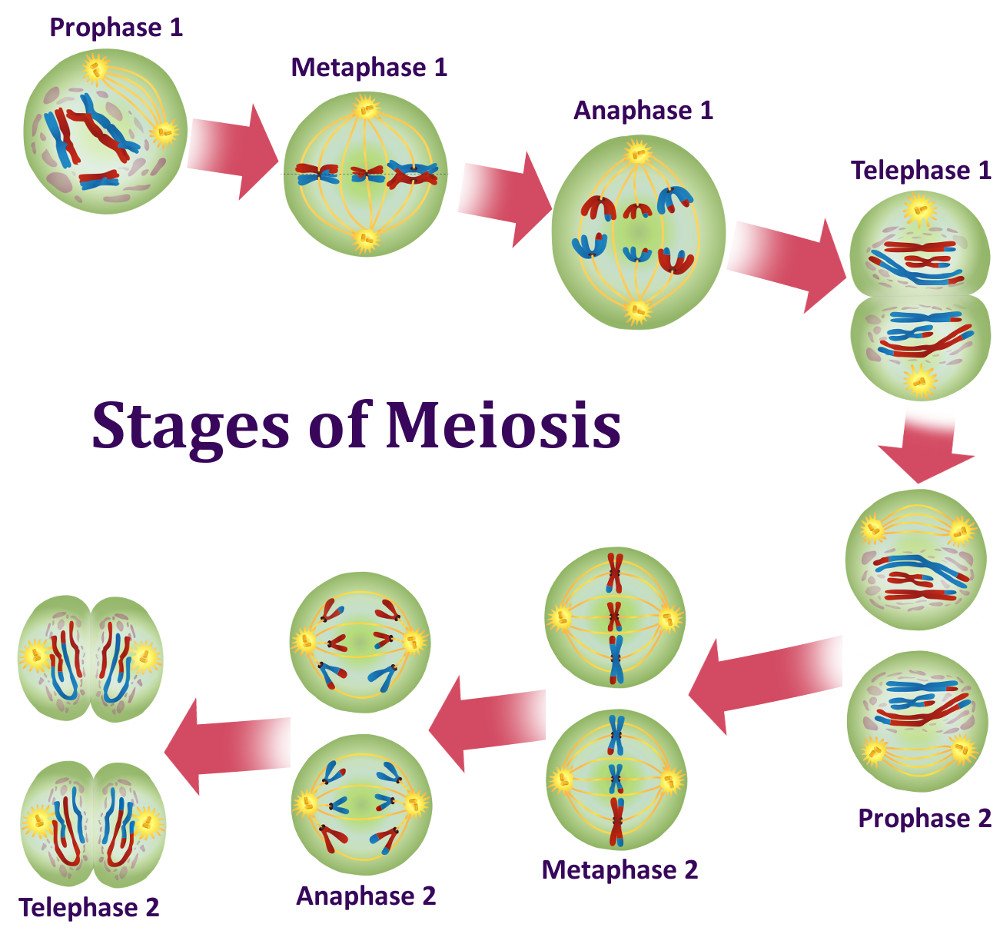
Mitotic Cell Division What Is Mitosis? What Is Meiosis?
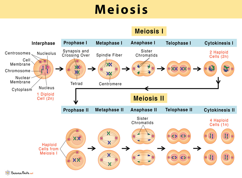
Meiosis Definition, Stages, & Purpose with Diagram

Meiosis, Stages, Meiosis vs Mitosis The Virtual Notebook

Cell Biology Glossary Meiosis Draw It to Know It
[Solved] Draw the process of Meiosis. Your parent cell is 2n=6 (n is
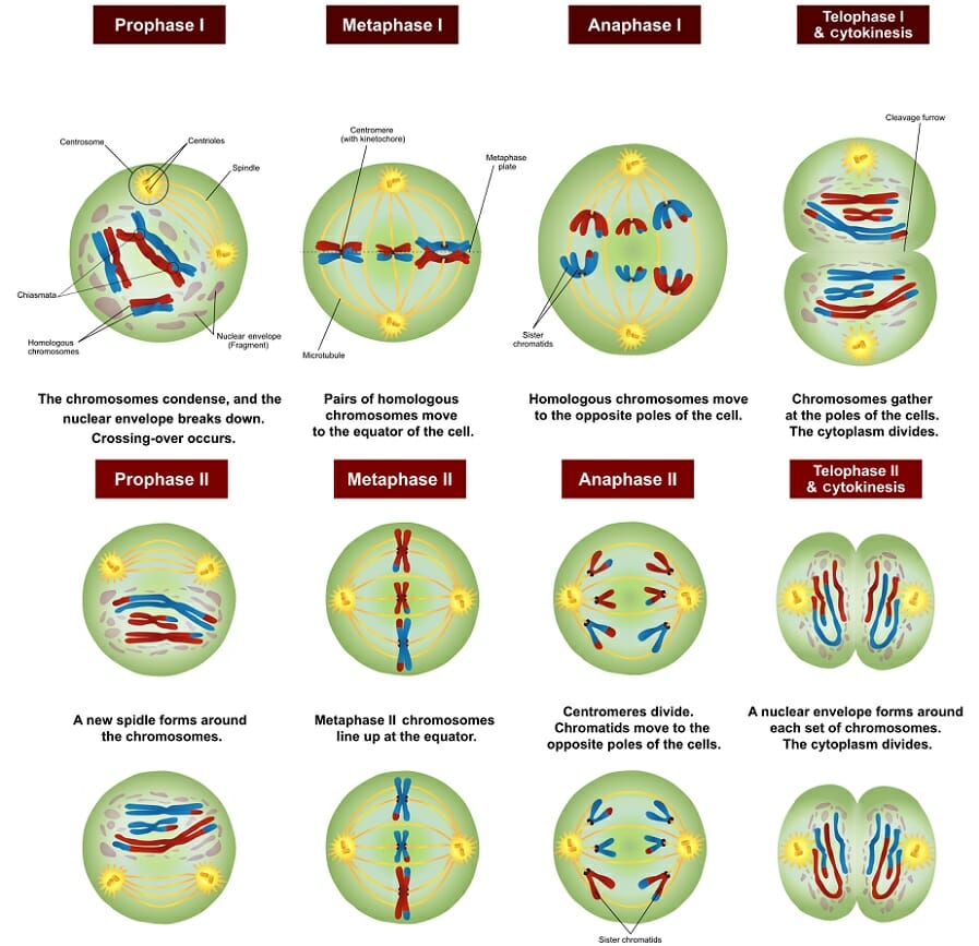
Meiosis Definition, Stages, Function and Purpose Biology Dictionary
Meiosis Phases, Stages, Applications with Diagram
Meiosis Phases Explore the various stages of meiosis

What is meiosis? Facts
Explain The Differences Between Meiosis And Mitosis.
This Means There Are 4 Copies Of Each Gene, Present In 2 Full Sets Of Dna, Each Set Having 2 Alleles.
Spindle Fibres Attach To The Chromosomes At The Centromere.
This Is The Well Labelled Diagram Of Meiosis.
Related Post: