Drawing Blood Vessels
Drawing Blood Vessels - Web peripheral artery disease: These can be drawn in transverse section (ts) and longitudinal section (ls) arteries are blood vessels that carry blood at high pressures away from the heart. 18k views 1 year ago class 7. Blood flow through the heart. A blown vein, sometimes called a ruptured vein, is a blood vessel that’s damaged due to a needle insertion. < prev next > 5 arterial blood sampling. When more than a few drops of blood are required, phlebotomists perform a venipuncture, typically of a surface vein in the arm. Web drawing blood vessels using illustrator’s blend tools by annie campbell — learn medical art. Blood vessels flow blood throughout the body. Great vessels of the heart. This condition affects the arteries in the legs and/or arms and may cause problems with wound healing and/or claudication (pain with movement, especially when walking).; The information given here supplements that given in chapters 2 and 3. Web peripheral artery disease: Disease of the arteries in the heart can predispose to blood clots, which may cause a heart attack. The. This can happen when a healthcare provider, such as a phlebotomist or nurse, draws blood or inserts a peripheral iv to give you medications or fluids. For adult patients, the most common and first choice is the median cubital vein in the antecubital fossa. Web describe the basic structure of a capillary bed, from the supplying metarteriole to the venule. Drawing blood usually isn’t taught in nursing school—more on why below—but it’s a critical skill for all types of nurses to learn. Base (posterior), diaphragmatic (inferior), sternocostal (anterior), and left and right pulmonary surfaces. When more than a few drops of blood are required, phlebotomists perform a venipuncture, typically of a surface vein in the arm. Discuss several factors affecting. Distinguish between elastic arteries, muscular arteries, and arterioles on the basis of structure, location, and function. The chapter includes background information (section 2.1), practical guidance (section 2.2) and illustrations (section 2.3) relevant to best practices in phlebotomy. Veins return blood back toward the heart. If you’ve ever had your blood drawn, you may have noticed veins on the inside of. Compare and contrast the three tunics that make up the walls of most blood vessels. Web by the end of this section, you will be able to: Drawing of blood vessel stock illustrations. Web plan diagrams show the structures of arteries and veins; The heart has five surfaces: Web this chapter covers all the steps recommended for safe phlebotomy and reiterates the accepted principles for blood drawing and blood collection (31). A blown vein, sometimes called a ruptured vein, is a blood vessel that’s damaged due to a needle insertion. Compare and contrast veins, venules, and venous sinuses on the basis of structure, location, and function. Blood vessels. Veins in your legs fight gravity to push blood up toward your heart. Web peripheral artery disease: Blood vessels flow blood throughout the body. When more than a few drops of blood are required, phlebotomists perform a venipuncture, typically of a surface vein in the arm. This can happen when a healthcare provider, such as a phlebotomist or nurse, draws. New 3d rotate and zoom. Web the most appropriate site to draw blood is selected based on vessel accessibility, patient age, and health status. Discuss several factors affecting blood flow in the venous system. Web describe the basic structure of a capillary bed, from the supplying metarteriole to the venule into which it drains. When more than a few drops. Compare and contrast veins, venules, and venous sinuses on the basis of structure, location, and function. New 3d rotate and zoom. Distinguish between elastic arteries, muscular arteries, and arterioles on the basis of structure, location, and function. Compare and contrast the three tunics that make up the walls of most blood vessels. 18k views 1 year ago class 7. Web drawing of blood vessel. 7.2k views 3 years ago scientific illustration | adobe illustrator. The three major types of blood vessels: A blown vein, sometimes called a ruptured vein, is a blood vessel that’s damaged due to a needle insertion. Web by the end of this section, you will be able to: Web this chapter covers all the steps recommended for safe phlebotomy and reiterates the accepted principles for blood drawing and blood collection (31). Compare and contrast the three tunics that make up the walls of most blood vessels. The information given here supplements that given in chapters 2 and 3. Web peripheral artery disease: Web blood, by definition, is a fluid that moves through the vessels of a circulatory system. Web drawing blood vessels using illustrator’s blend tools by annie campbell — learn medical art. Great vessels of the heart. Blood vessels flow blood throughout the body. Web learn all about the heart, blood vessels, and composition of blood itself with our 3d models and explanations of cardiovascular system anatomy and physiology. Capillaries surround body cells and tissues to deliver and absorb oxygen, nutrients, and other substances. These can be drawn in transverse section (ts) and longitudinal section (ls) arteries are blood vessels that carry blood at high pressures away from the heart. Some are big enough to see under your skin. Disease of the arteries in the heart can predispose to blood clots, which may cause a heart attack. When more than a few drops of blood are required, phlebotomists perform a venipuncture, typically of a surface vein in the arm. The first step in drawing blood correctly is to identify the appropriate veins to puncture. Drawing blood usually isn’t taught in nursing school—more on why below—but it’s a critical skill for all types of nurses to learn.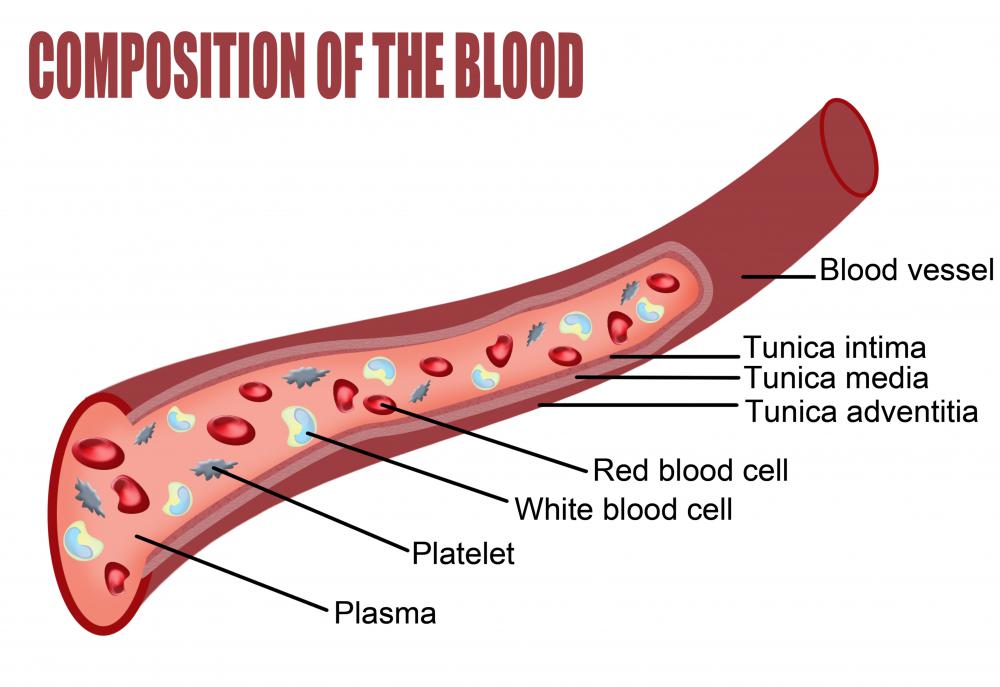
What are Blood Vessels? (with pictures)
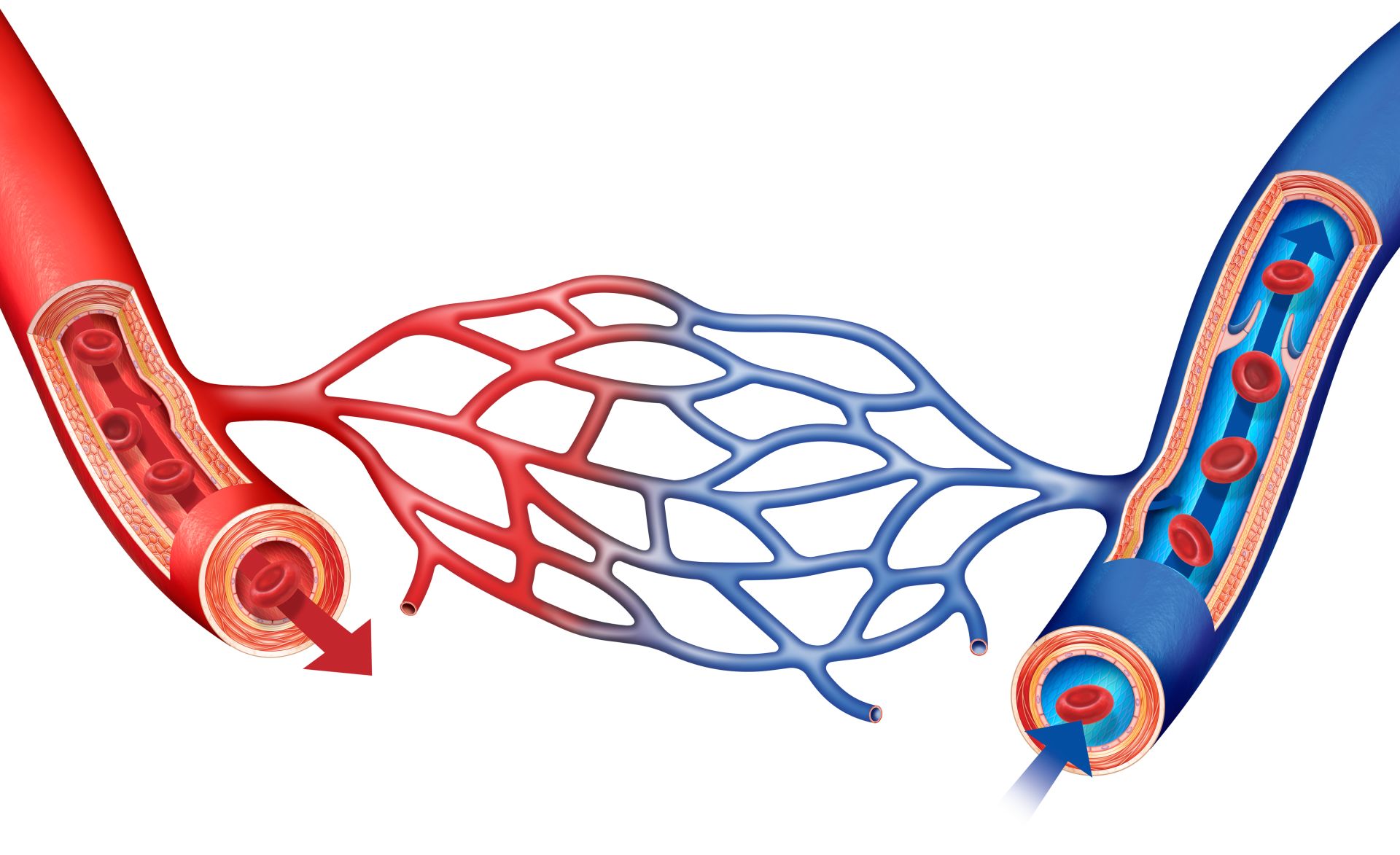
What Are Blood Vessels Blood Vessel Facts DK Find Out
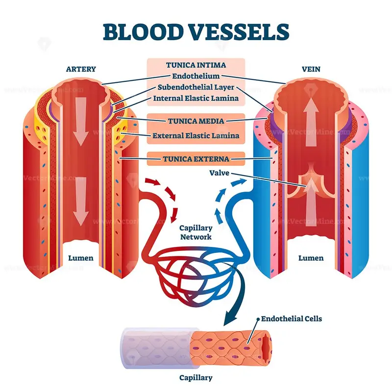
Blood vessels with artery and vein internal structure vector

What are the 5 types of blood vessels?

Blood vessel Definition, Anatomy, Function, & Types Britannica
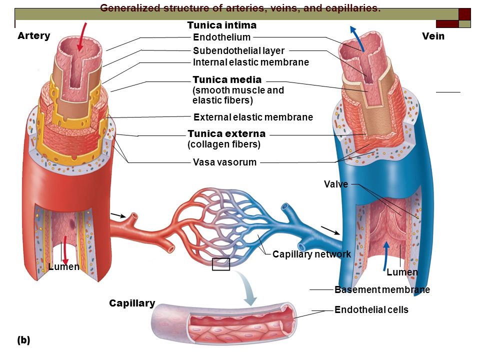
Blood Vessels Alisa Houghton

How to draw Structure of heart and blood circulation drawing step by
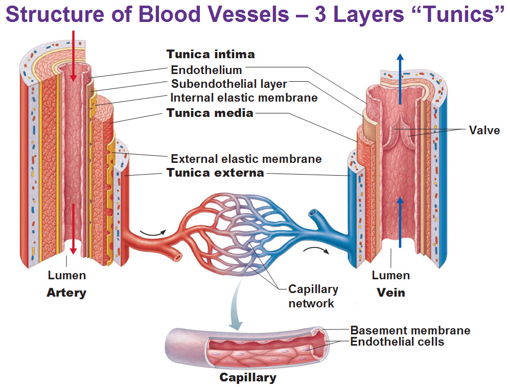
Blood vessels (Types, structure and functions) Online Science Notes
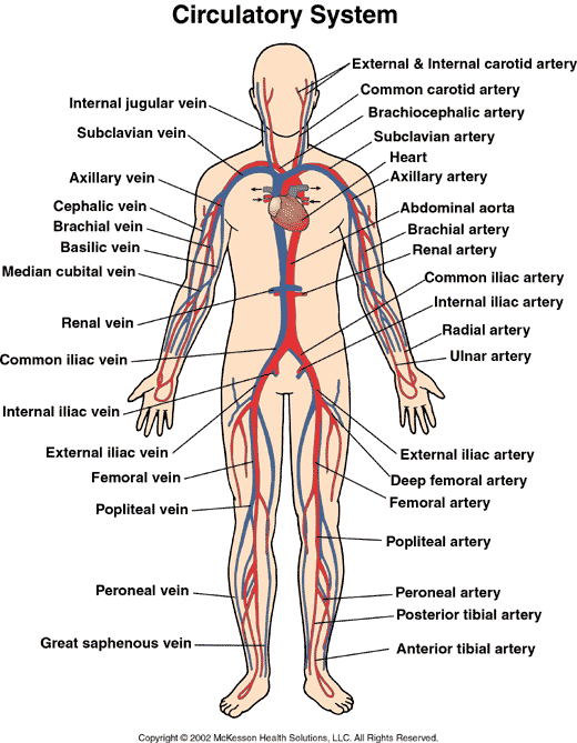
What are the Three Types of Blood Vessels and Their Functions? First
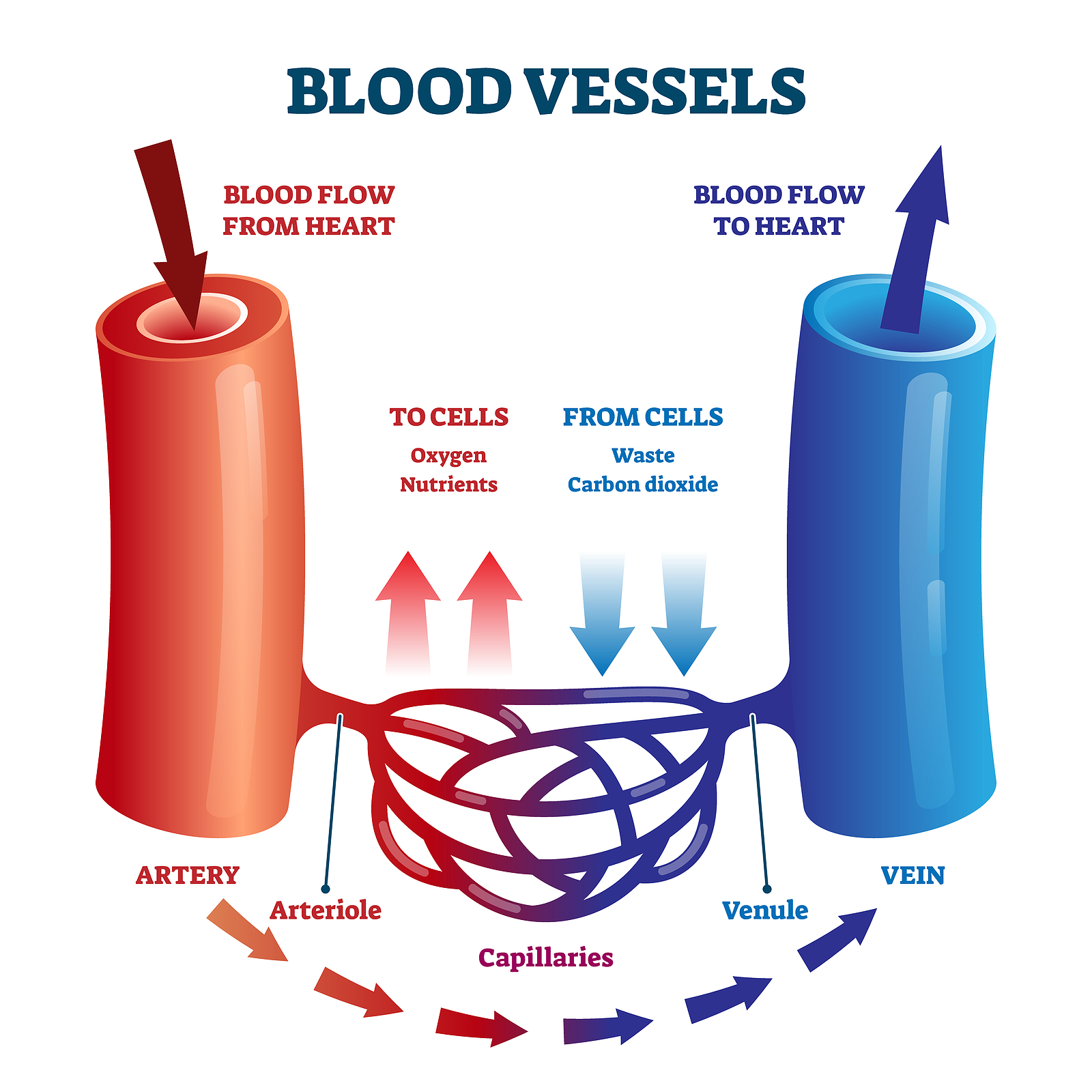
How blood flows through the body MooMooMath and Science
View Drawing Of Blood Vessel Videos.
Erythrocytes (Red Blood Cells) Leukocytes (White Blood Cells) Thrombocytes (Platelets) Clinical Notes.
The Heart Has Five Surfaces:
< Prev Next > 5 Arterial Blood Sampling.
Related Post: