Drawing Of The Spinal Cord
Drawing Of The Spinal Cord - The spinal cord is divided into five different parts. I am creating these videos to further supplement the virtual learning of other medical. However, the following arteries branch from the vertebral arteries to directly supply the spinal cord itself: Your spinal cord is a cylindrical structure that runs through the center of your spine, from your brainstem to your low back. 94k views 4 years ago. It forms a vital link between the brain and the body. The spinal cord is a cylindrical mass of neural tissue extending from the caudal aspect of the medulla oblongata of the brainstem to the level of the first lumbar vertebra (l1). The spinal cord is a long bundle of nerves and cells that carries signals between the. Blood supply of the spinal cord. Web anatomy of the spinal cord. The spinal cord is part of the central nervous system (cns), which extends caudally and is protected by the bony structures of the vertebral column. Ventral funiculus, cuneate fasciculus, gracile fasciculus. View spinal cord drawing videos. 160k views 12 years ago. Let us learn how to draw a. Representation in 3/4 front view of the stucture of the spinal cord, and rachidian nerves. Spine isolated on a white backgrounds. Let us learn how to draw a. Web spine anatomy, diagram & pictures | body maps. Your spinal cord is a cylindrical structure that runs through the center of your spine, from your brainstem to your low back. The nervous system is divided into two main parts: It is covered by the three membranes of the cns, i.e., the dura mater, arachnoid and the innermost pia mater. From the spinal cord se dã©tachent on each side rachidian nerves constituted of a ganglion, a posterior and an anterior root. It then travels inferiorly within the vertebral canal, surrounded by. 94k views 4 years ago. Web keep learning about the white and grey matter of the spinal cord using our spinal cord diagram labeling exercises and quizzes! From the spinal cord se dã©tachent on each side rachidian nerves constituted of a ganglion, a posterior and an anterior root. The spinal cord is divided into five different parts. Web this article. Hand drawn spine isolated on a white backgrounds. Web spinal cord, drawing the spinal cord. Spine isolated on a white backgrounds. Blood vessels of the spinal cord [12:23] arteries and veins of the spinal cord. Web dabrafenib with trametinib can halt growth of some tumours for more than three times as long as standard chemotherapy, study shows Ventral funiculus, cuneate fasciculus, gracile fasciculus. What is the spinal cord? Web how to draw t.s. I am creating these videos to further supplement the virtual learning of other medical. View spinal cord drawing videos. Web overview of spinal cord anatomy. The spinal cord is part of the central nervous system (cns), which extends caudally and is protected by the bony structures of the vertebral column. The spinal cord is part of the central nervous system and consists of a tightly packed column of nerve tissue that extends downwards from the brainstem through the central. Spine isolated on a white backgrounds. I am creating these videos to further supplement the virtual learning of other medical. Blood supply of the spinal cord. Web keep learning about the white and grey matter of the spinal cord using our spinal cord diagram labeling exercises and quizzes! In adults, the spinal cord is usually 40cm long and 2cm wide. Representation in 3/4 front view of the stucture of the spinal cord, and rachidian nerves. Blood supply of the spinal cord. However, the following arteries branch from the vertebral arteries to directly supply the spinal cord itself: The spinal cord is a cylindrical mass of neural tissue extending from the caudal aspect of the medulla oblongata of the brainstem to. The spinal cord is part of the central nervous system and consists of a tightly packed column of nerve tissue that extends downwards from the brainstem through the central column of the spine. Spinal cord drawing stock vectors. Let us learn how to draw a. Web how to draw t.s. The spinal cord is part of the central nervous system. The central nervous system, consisting of the brain and spinal cord, and the peripheral nervous system, made up of nerves and ganglia. Web anatomy of the spinal cord. Web in this video i will guide you through the creation of your own drawing of a spinal cord cross section. Web choose from drawing of spinal cord stock illustrations from istock. View spinal cord drawing videos. Spine isolated on a white backgrounds. Web the incidence of spinal metastases (sm) is reported to be around 16% in solid tumors and result in meaningful impairment of quality of life (qol) due to pain or neurological deficits [1,2,3,4].the current treatment of sm involves spinal surgery and adjuvant radiotherapy (rt) and systemic therapy (ctx) [].surgery aims to protect the. Web how to draw t.s. The nervous system is divided into two main parts: 94k views 4 years ago. Web dabrafenib with trametinib can halt growth of some tumours for more than three times as long as standard chemotherapy, study shows Hand drawn spine isolated on a white backgrounds. It's a delicate structure that contains nerve bundles and cells that carry messages from your brain to the rest of your body. It is covered by the three membranes of the cns, i.e., the dura mater, arachnoid and the innermost pia mater. Let us learn how to draw a. From the spinal cord se dã©tachent on each side rachidian nerves constituted of a ganglion, a posterior and an anterior root.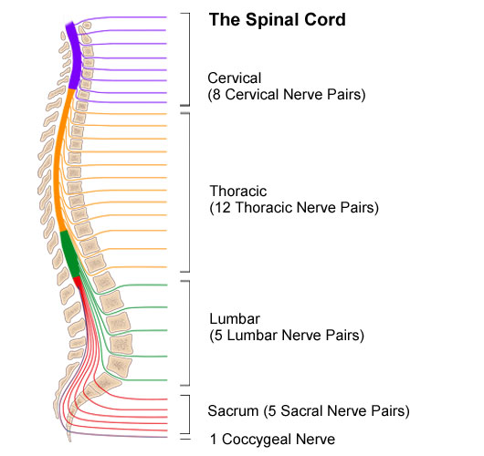
Anatomy of the Spinal Cord Stanford Medicine Children's Health

The Spinal Cord Neurologic Clinics
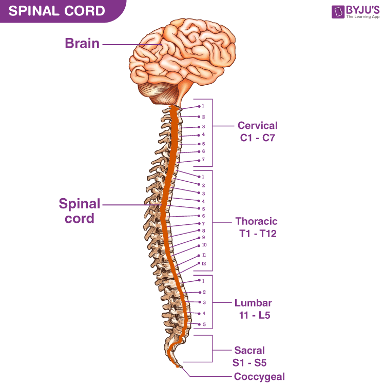
Spinal Cord Anatomy, Structure, Function, & Diagram

How to Draw Structure Of The Spinal Cord Diagram Easy And Step by Step

Spinal Cord Drawing at GetDrawings Free download
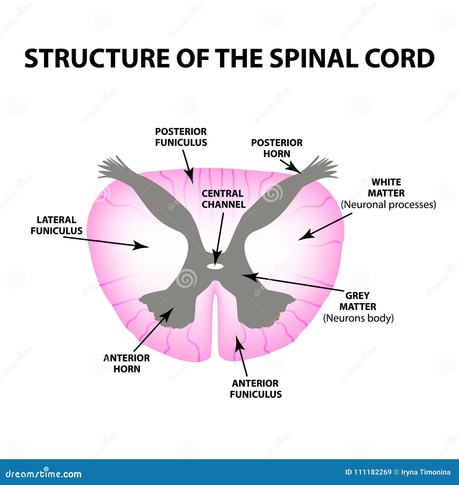
The Structure of the Spinal Cord. Infographics. Vector Illustration on
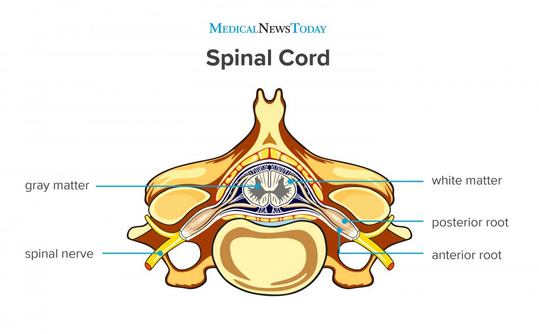
Spinal cord Anatomy, functions, and injuries
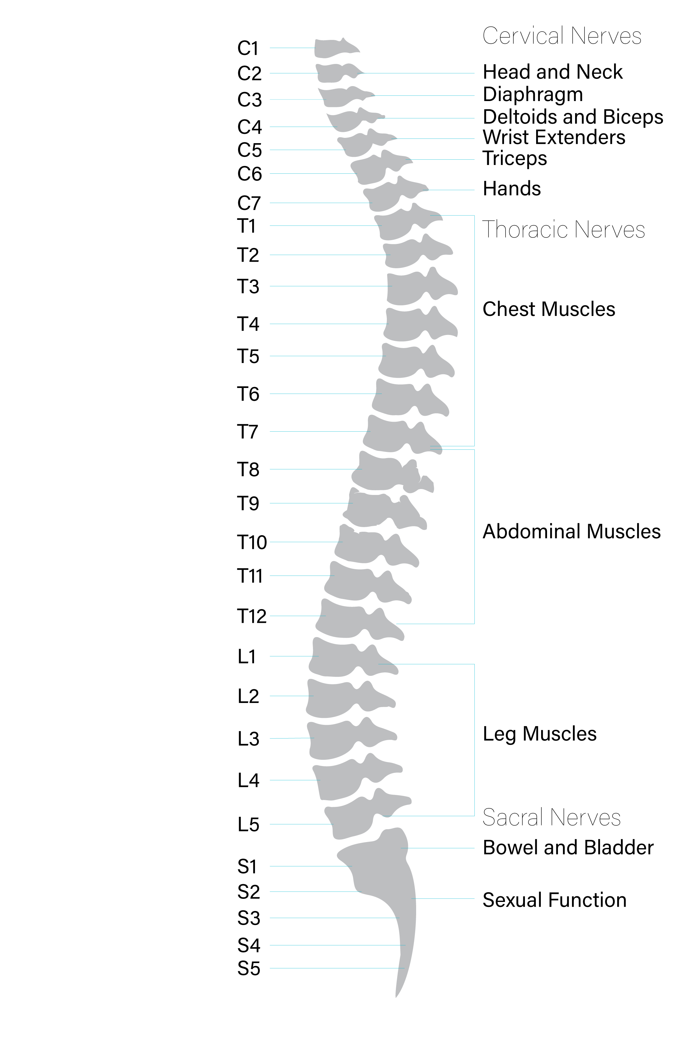
Anatomy of the Spinal Cord Praxis Spinal Cord Institute

Spinal Cord, Drawing Stock Image C017/1520 Science Photo Library

How To Draw Spinal Cord Step By Step Easily YouTube
What Is The Spinal Cord?
Ventral Funiculus, Cuneate Fasciculus, Gracile Fasciculus.
Spinal Cord, Funiculi Of Spinal Cord, Tectospinal Tract, Anterior Funiculus;
The Spinal Cord Is A Cylindrical Mass Of Neural Tissue Extending From The Caudal Aspect Of The Medulla Oblongata Of The Brainstem To The Level Of The First Lumbar Vertebra (L1).
Related Post: