Female Reproductive Anatomy Drawing
Female Reproductive Anatomy Drawing - The function of your external genitals are to protect the internal parts from infection and allow sperm to enter your vagina. Female reproductive anatomy diagram drawing. Anatomy of the female reproductive system; Web female anatomy includes the internal and external reproductive organs. Female reproductive system of internal organs continuous line. External structures include the mons pubis, pudendal cleft, labia majora and minora, vulva, bartholin’s gland, and the clitoris. The vulva refers to the external female genitalia. The internal female sex organs form a pathway, the internal female genital tract, composed of the vagina, uterus, the paired uterine tubes and ovaries. Female reproductive organs undergo substantial structural and functional changes every month. The female reproductive anatomy includes both external and internal parts. Female reproductive organs undergo substantial structural and functional changes every month. Your vulva is the collective name for all your external genitals. Web browse 1,300+ anatomy of the female reproductive system drawing stock photos and images available, or start a new search to explore more stock photos and images. View female reproductive anatomy diagram drawing videos. Web these are the. The components of the external female genitalia occupy a large part of the female perineum and collectively form what's known as the vulva. The female gonads, the ovaries produce ova. Web female anatomy includes the internal and external reproductive organs. Egg cell production, or oogenesis, begins with the primordial follicles. External structures include the mons pubis, pudendal cleft, labia majora. After that time, fertility declines more rapidly, until it ends completely at the end of menopause. This article provides diagrams with supporting information to help you learn about the main structures and functions. Egg cell production, or oogenesis, begins with the primordial follicles. It also produces female sex hormones,. The vagina, uterus, ovaries and uterine tubes compose the internal genital. The vagina is part of the internal genitalia of the female reproductive system. Anatomy and physiology of the female reproductive system. Female reproductive organs undergo substantial structural and functional changes every month. The vagina, uterus, ovaries and uterine tubes compose the internal genital organs. View female reproductive anatomy diagram drawing videos. The female gonads, the ovaries produce ova. They produce oocytes (egg cells), as well as estrogen, progesterone, and other hormones. Web design by diego sabogal. The female reproductive anatomy includes both external and internal parts. Web female anatomy includes the internal and external reproductive organs. Female fertility (the ability to conceive) peaks when women are in their twenties, and is slowly reduced until a women reaches 35 years of age. Female reproductive organs undergo substantial structural and functional changes every month. These organs are supported in the pelvis by ligaments. Web female anatomy includes the internal and external reproductive organs. Anatomy of the female reproductive. Mons pubis, labia majora, labia minora, clitoris, vestibule, hymen, vestibular bulb and vestibular glands. Web the female reproductive tract is all located within the pelvis. Cup internal genitals of women. Web these are the mons pubis, labia majora and minora, clitoris, vestibule, vestibular bulb and glands. Female reproductive system of internal organs continuous line. An female’s internal reproductive organs are the vagina, uterus, fallopian tubes, cervix, and ovary. The vulva refers to the external female genitalia. Web choose from female reproductive anatomy drawings stock illustrations from istock. External structures include the mons pubis, pudendal cleft, labia majora and minora, vulva, bartholin’s gland, and the clitoris. However, it also has the additional task of supporting. Web what are the parts of the female reproductive system? Cup internal genitals of women. Your vulva is the collective name for all your external genitals. Mons pubis, labia majora, labia minora, clitoris, vestibule, hymen, vestibular bulb and vestibular glands. Web female anatomy includes the external genitals, or the vulva, and the internal reproductive organs. An female’s internal reproductive organs are the vagina, uterus, fallopian tubes, cervix, and ovary. The vagina serves a multitude of functions. Web the female reproductive system functions to produce gametes and reproductive hormones, just like the male reproductive system; Ovaries are the female gonads. Web browse 450+ female reproductive anatomy drawing stock photos and images available, or start a new. Anatomy of the female reproductive system; Web female anatomy includes the external genitals, or the vulva, and the internal reproductive organs. Female reproductive organs undergo substantial structural and functional changes every month. Learn all about fertilization, pregnancy, delivery, and lactation. Female reproductive anatomy diagram drawing. The internal female sex organs form a pathway, the internal female genital tract, composed of the vagina, uterus, the paired uterine tubes and ovaries. Cup internal genitals of women. These organs are supported in the pelvis by ligaments. This article provides diagrams with supporting information to help you learn about the main structures and functions. Web what are the parts of the female reproductive system? 3d models, illustrations, and accompanying descriptions guide you in exploring the anatomy and physiology of the female reproductive organs. Web these are the mons pubis, labia majora and minora, clitoris, vestibule, vestibular bulb and glands. Ovaries are the female gonads. Web design by diego sabogal. An female’s internal reproductive organs are the vagina, uterus, fallopian tubes, cervix, and ovary. The vagina, uterus, ovaries and uterine tubes compose the internal genital organs.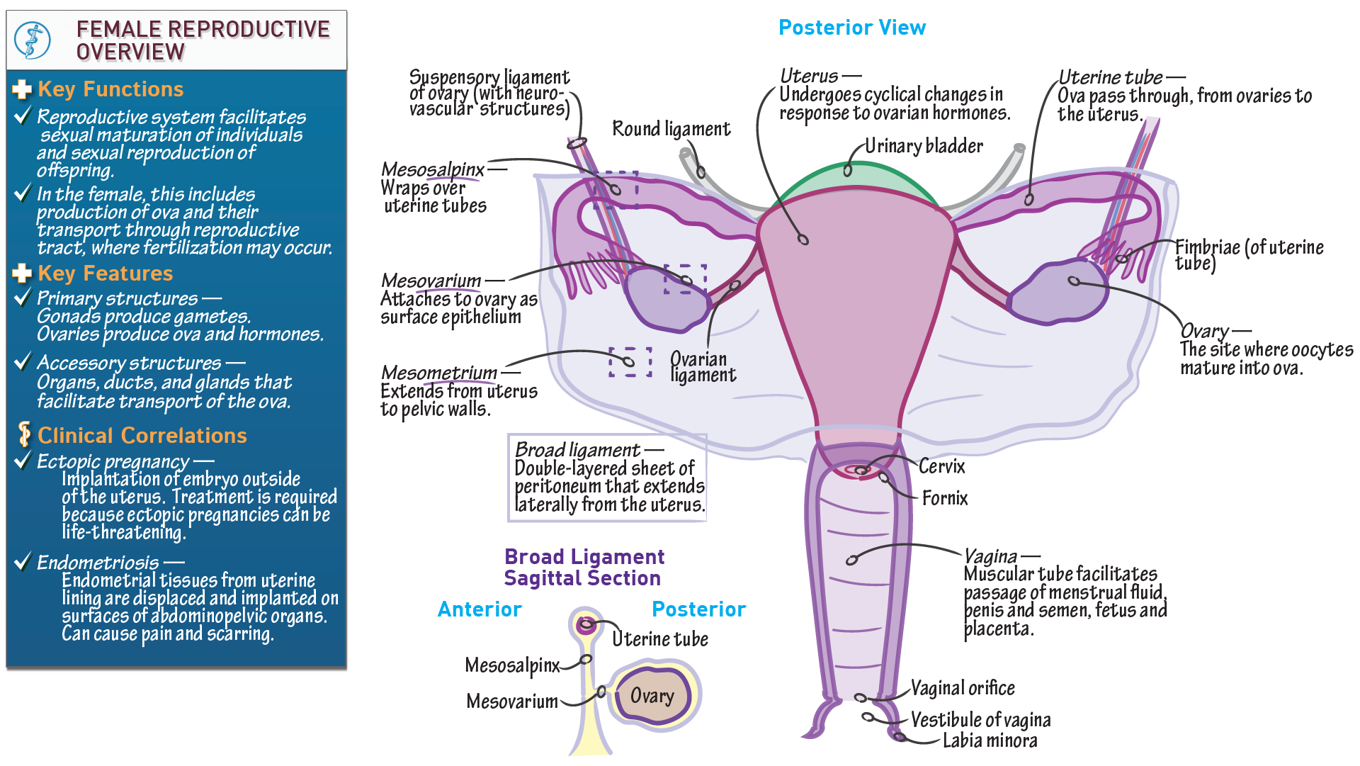
Anatomy & Physiology Anatomical Overview of the Female Reproductive

How to draw Female Reproductive system easily Step by step YouTube
.jpg?response-content-disposition=attachment)
Female Reproductive System resource Imageshare
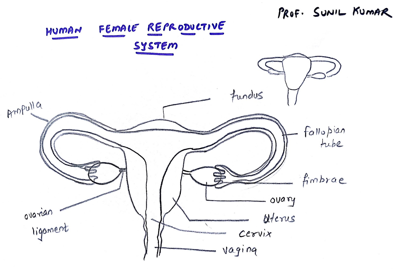
PROF. SUNIL KUMAR'S ADDABIOLOGY EASY WAY TO DRAW FEMALE REPRODUCTIVE

How to Draw Female Reproductive System, Diagram, Functions, Anatomy
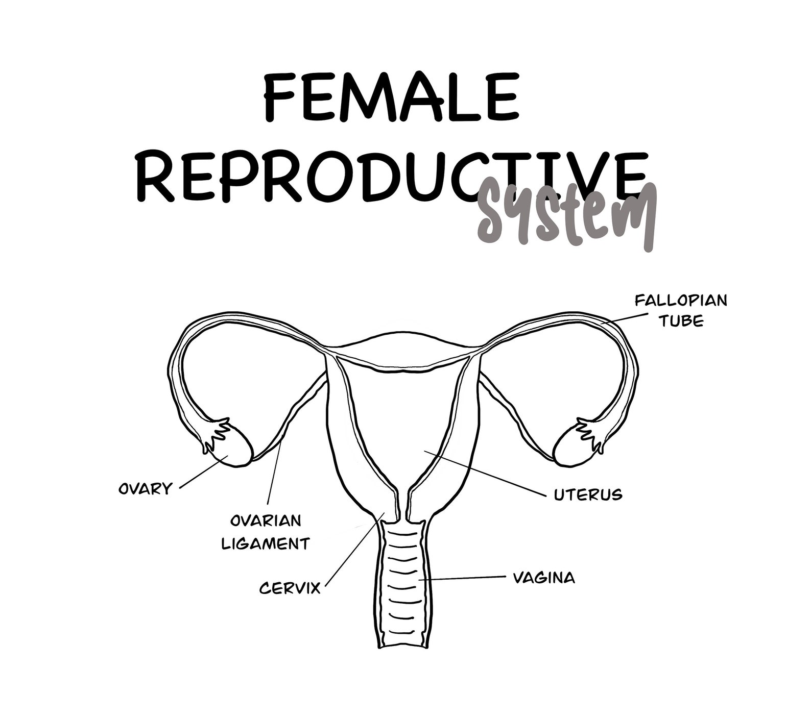
Female Reproductive System Educational Printable Etsy
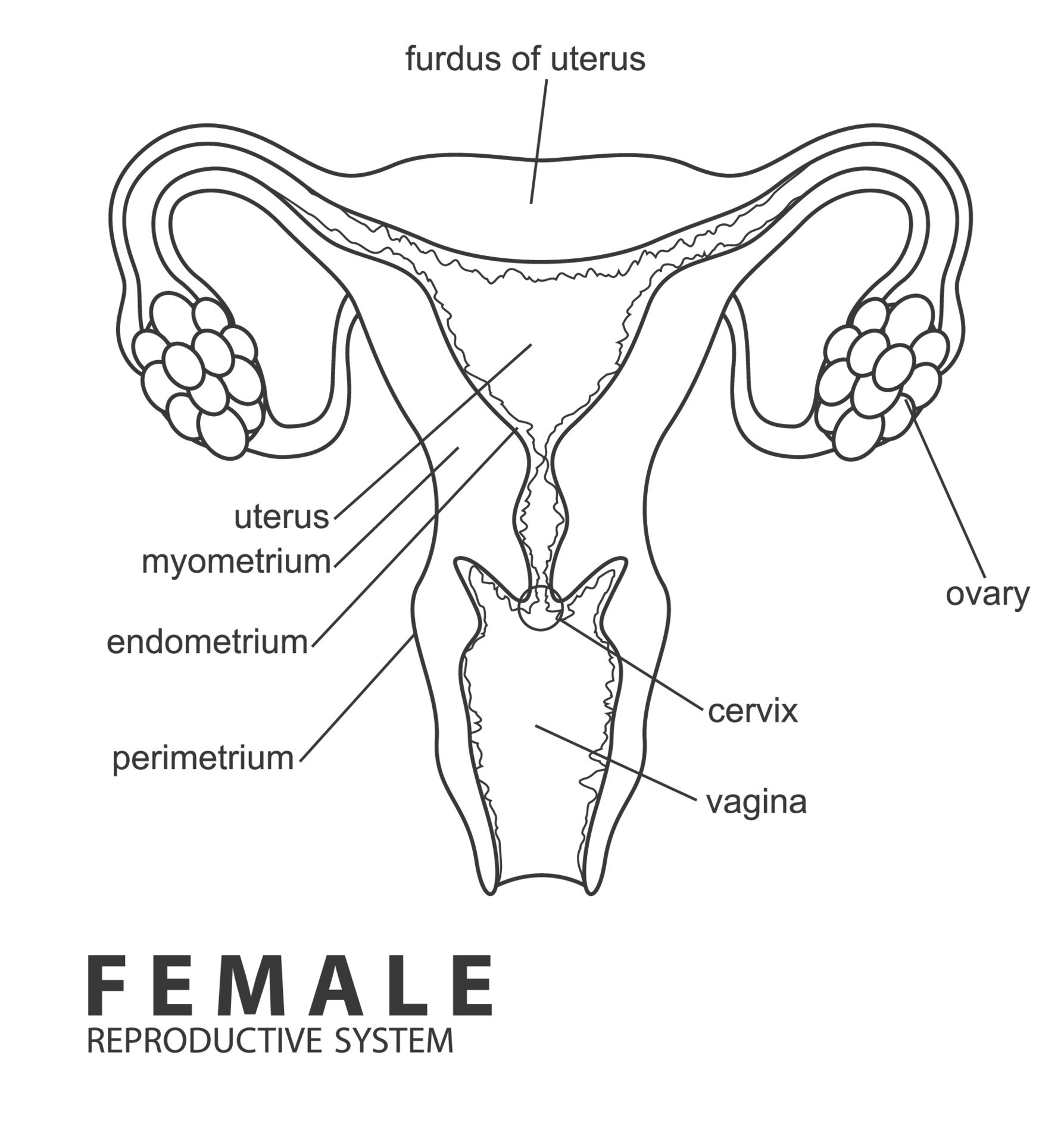
Female reproductive system outline, Vector Illustration 22674066 Vector
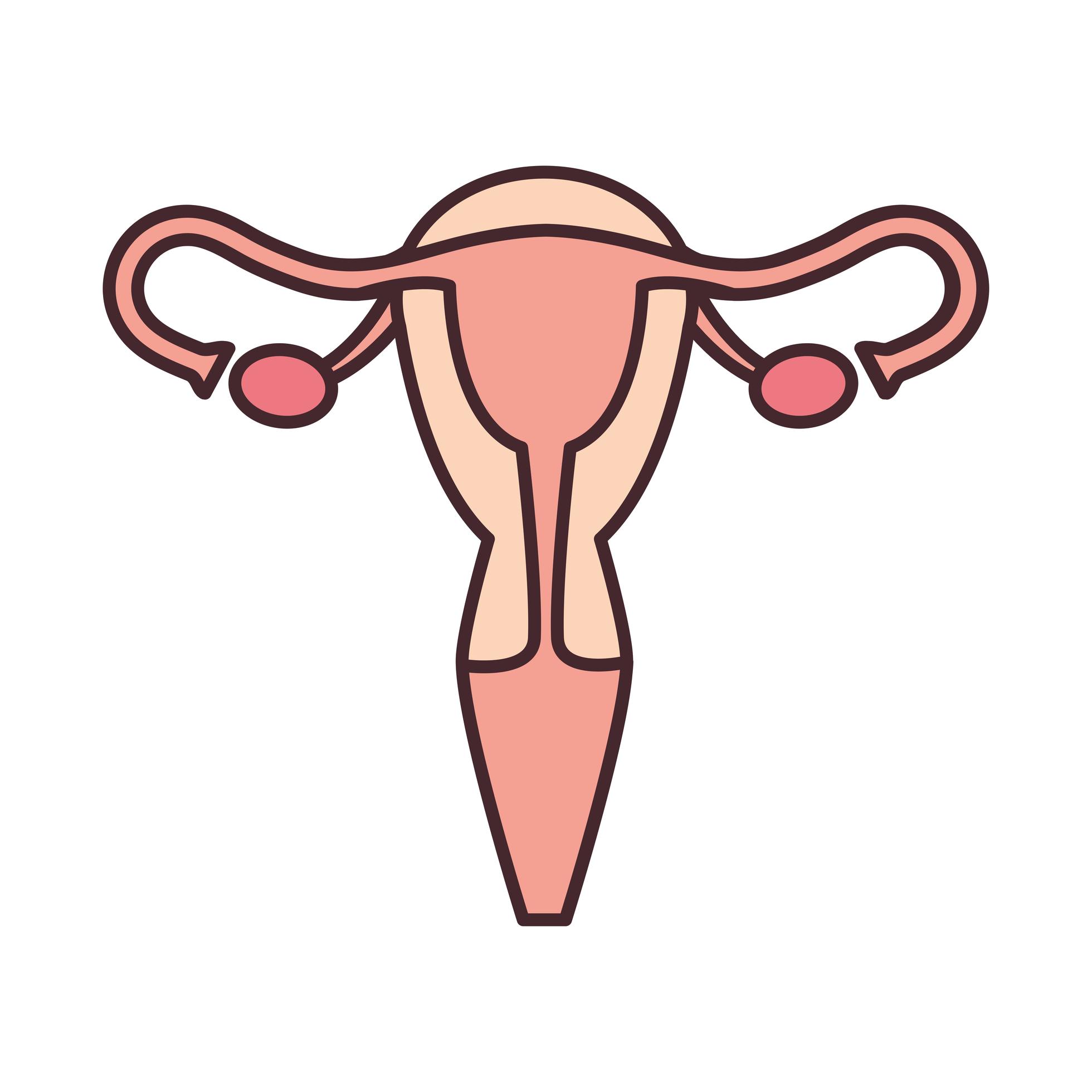
female reproductive system 2495597 Vector Art at Vecteezy
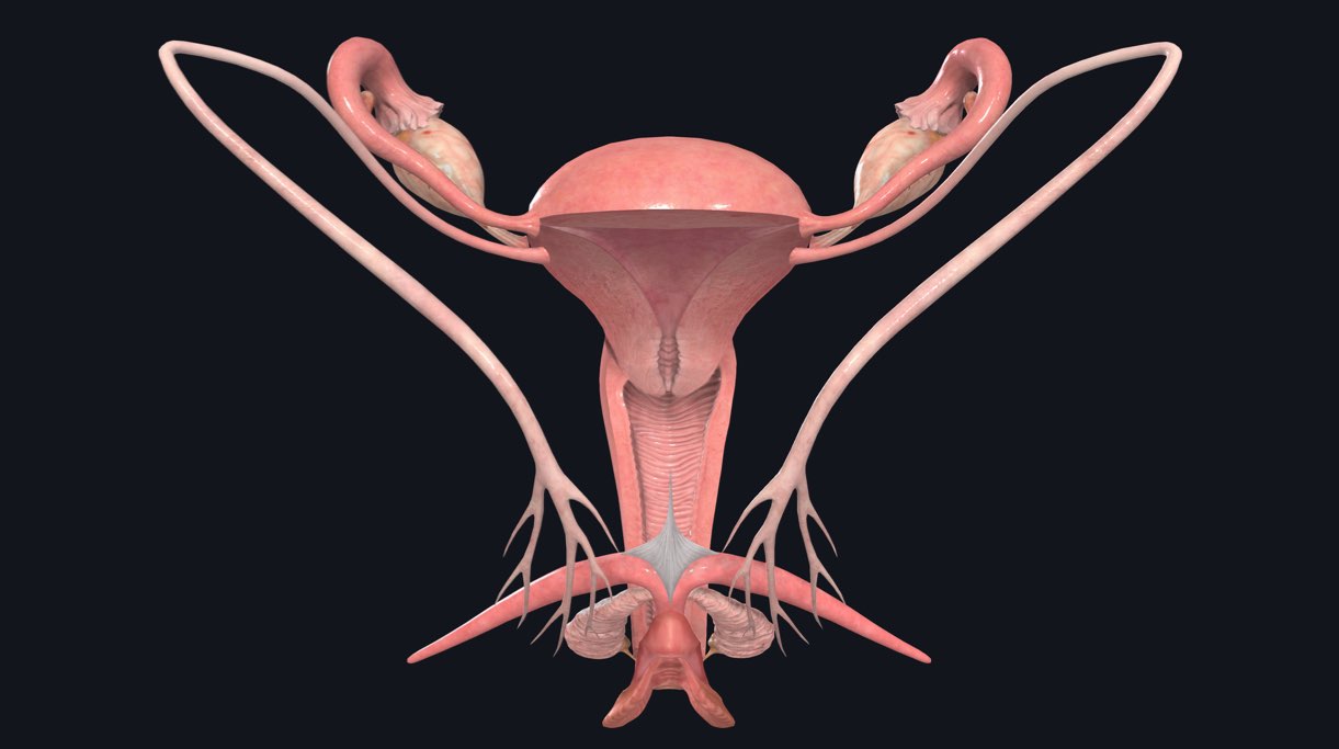
The female reproductive system Complete Anatomy

How to draw female reproductive system easily step by step YouTube
External Structures Include The Mons Pubis, Pudendal Cleft, Labia Majora And Minora, Vulva, Bartholin’s Gland, And The Clitoris.
After That Time, Fertility Declines More Rapidly, Until It Ends Completely At The End Of Menopause.
Female Fertility (The Ability To Conceive) Peaks When Women Are In Their Twenties, And Is Slowly Reduced Until A Women Reaches 35 Years Of Age.
Mons Pubis, Labia Majora, Labia Minora, Clitoris, Vestibule, Hymen, Vestibular Bulb And Vestibular Glands.
Related Post: