Microscope Parts Drawing
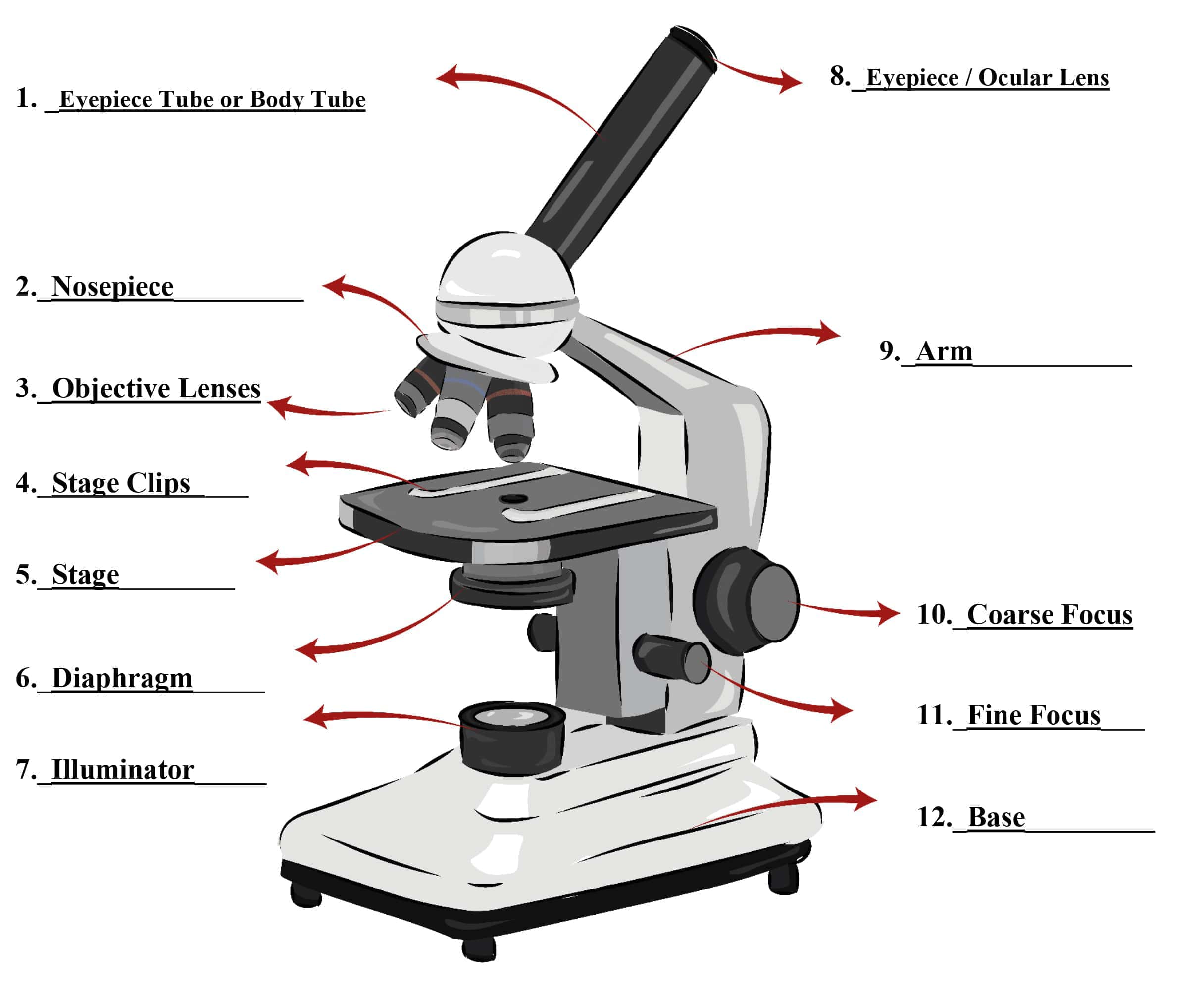
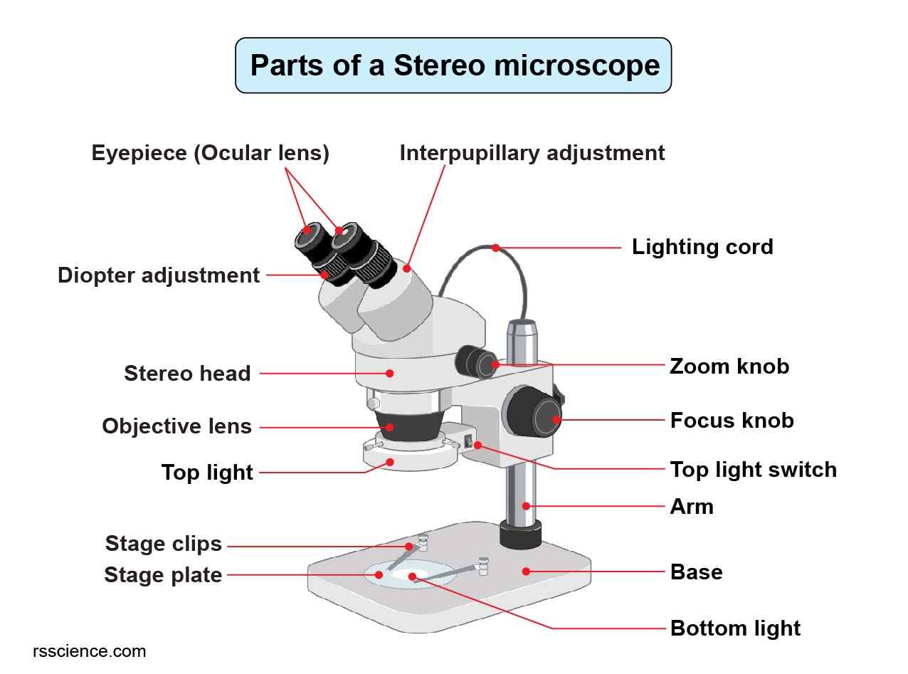
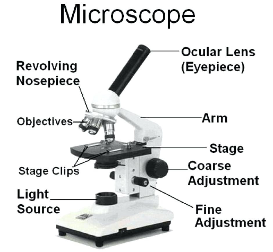
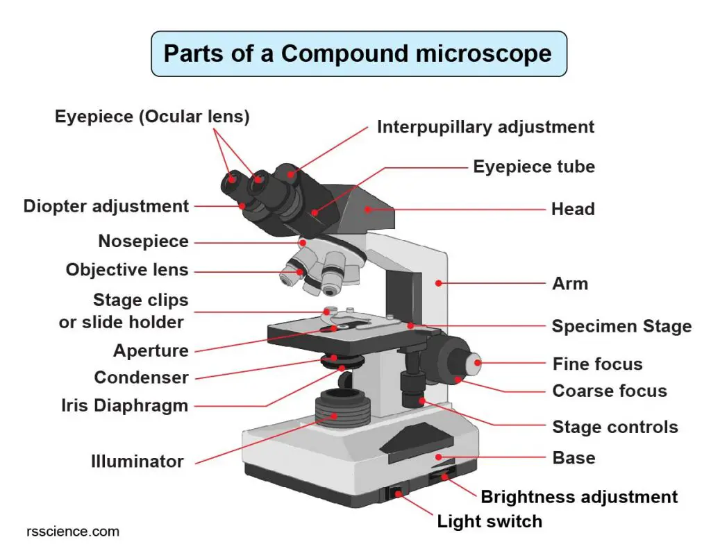
Microscope Parts Drawing - Web compound microscope parts labeled diagram. Web a microscope is an optical instrument used to magnify an image of a tiny object; Web microscope parts labeled. 📏 the microscope has three major structural parts: Diagram of parts of a microscope. Eyepiece (ocular lens) with or without pointer: The head, the base, and the arm. First form the body of the microscope. A microscope is one of the invaluable tools in the laboratory setting. Compound microscope definitions for labels. There are three major structural parts of a microscope: An object glass (objective) closer to the object or specimen, and an eyepiece (ocular) closer to the observer's eye (with means of adjusting the position of the specimen and the microscope lenses). Web a microscope is an instrument that magnifies objects otherwise too small to be seen, producing an image in. Useful as a means to change focus on one eyepiece so as to correct for any difference in vision between your two eyes. Web learn about the different parts of the microscope, including the simple microscope and the compound microscope, with labeled pictures and detailed explanations. Web without these components working in perfect harmony, scientific discoveries ranging from studying cells. Knobs (fine and coarse) 6. Most photographs of cells are taken using a microscope, and these pictures can also be called micrographs. Web learn about the different parts of the microscope, including the simple microscope and the compound microscope, with labeled pictures and detailed explanations. 📐 adjustment knobs are used to adjust the focus of the microscope. A microscope is. Web learn about the different parts of the microscope, including the simple microscope and the compound microscope, with labeled pictures and detailed explanations. Knobs (fine and coarse) 6. The part that is looked through at the top of the compound microscope. A microscope is a laboratory instrument used to examine objects that are too small to be seen by the. The lens the viewer looks through to see the specimen. The eyepiece usually contains a 10x or 15x power lens. A microscope is one of the invaluable tools in the laboratory setting. Compound microscope definitions for labels. Most photographs of cells are taken using a microscope, and these pictures can also be called micrographs. There are three structural parts of the microscope i.e. The most familiar kind of microscope is the optical microscope,. An object glass (objective) closer to the object or specimen, and an eyepiece (ocular) closer to the observer's eye (with means of adjusting the position of the specimen and the microscope lenses). Had a mountain of just computer parts and chips. Web microscope parts labeled. Eyepiece lens (ocular lens) and eyepiece tube. Web what are the parts of a microscope? The body tube connects the eyepiece to the objective lenses. Web without these components working in perfect harmony, scientific discoveries ranging from studying cells to examining microorganisms would not be possible. The head, the base, and the arm. The optical components contained within modern microscopes are mounted on a stable, ergonomically designed base that allows rapid exchange, precision centering, and careful alignment between those assemblies that are optically interdependent. Use this with the microscope parts activity to help students identify and label the main parts of a microscope and then describe. Web the 16 core parts of a compound microscope are: 📐 adjustment knobs are used to adjust the focus of the microscope. Web learn about the different parts of the microscope, including the simple microscope and the compound microscope, with labeled pictures and detailed explanations. Web the goal is to complete a drawing of a microscope by creating each part. Eyepiece (ocular lens) with or without pointer: The common types of microscopes are: The width of a human hair and can't be seen without a microscope. Web the goal is to complete a drawing of a microscope by creating each part one part at a time. There are three major structural parts of a microscope: It will take 9 steps in total to complete the drawing. Eyepiece lens (ocular lens) and eyepiece tube. The common types of microscopes are: Web compound microscope parts labeled diagram. This article delves into the symphony of microscope parts, exploring how each component plays a vital role in scientific discovery. Diagram of parts of a microscope. 450 views 3 years ago #chatgpt #drawing #microscope. Connects the eyepiece to the objective lenses. Web without these components working in perfect harmony, scientific discoveries ranging from studying cells to examining microorganisms would not be possible. What is the difference between compound microscope and simple. Here are some key points: Ready to take your drawing skills to the next level? Web what are the parts of a microscope? In its simplest form, the compound microscope consisted of two convex lenses aligned in series: The eyepiece usually contains a 10x or 15x power lens. The finished drawing will be embellished with a tad bit of color making it a drawing you will be proud to show off!
Parts of a Microscope SmartSchool Systems

5 Types of Microscopes with Definitions, Principle, Uses, Labeled Diagrams

Parts Of A Microscope With Functions And Labeled Diagram Images

How to Use a Microscope

Parts of a microscope with functions and labeled diagram

Compound Microscope Parts, Functions, and Labeled Diagram New York

301 Moved Permanently

Parts Of A Microscope With Functions And Labeled Diagram Images

Compound Microscope Parts Labeled Diagram and their Functions Rs

Labeled Microscope Diagram Tim's Printables
It Is Used To Observe Things That Cannot Be Seen By The Naked Eye.
A Microscope Is A Laboratory Instrument Used To Examine Objects That Are Too Small To Be Seen By The Naked Eye.
The Lens The Viewer Looks Through To See The Specimen.
A Microscope Is One Of The Invaluable Tools In The Laboratory Setting.
Related Post: